Tuesday Poster Session
Category: IBD
P4435 - Endometriosis Implantation Masquerading as Stricturing Crohn’s Disease of the Terminal Ileum: A Case Report
Tuesday, October 29, 2024
10:30 AM - 4:00 PM ET
Location: Exhibit Hall E

Has Audio

Puja Sasankan, BS
George Washington University School of Medicine and Health Sciences
Washington, DC
Presenting Author(s)
Puja Sasankan, BS1, Vinay Rao, MD1, Marie L.. Borum, MD, MPH, FACG1, Samuel A.. Schueler, MD1, Natasha Kamal, MD2
1George Washington University School of Medicine and Health Sciences, Washington, DC; 2Frederick Gastroenterology Associates, Frederick, MD
Introduction: Crohn’s disease (CD) is characterized by transmural inflammation which may involve the entire gastrointestinal tract, most commonly the terminal ileum (TI). Endometriosis (EM) is the ectopic implantation of endometrial glands and stroma in locations outside the uterus, including the bowel (most commonly the left colon). Symptoms of EM affecting the bowel mimic CD and include abdominopelvic pain, constipation or diarrhea, dysmenorrhea, dyschezia, and dyspareunia. We present a rare case of endometrial bowel implantation leading to TI stricturing.
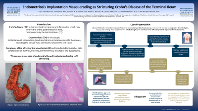
Case Description/Methods: A 38-year-old female with EM with history of hysterectomy and cholecystectomy presented with one year of progressive abdominal pain, nausea, and vomiting thought secondary to EM and initially improved after hysterectomy. Symptoms recurred months later prompting hospital admission where she was found to have mild leukocytosis, mild C-reactive protein (CRP) and erythrocyte sedimentation rate (ESR) elevation, normal fecal calprotectin, and computed tomography (CT) noting irregular enhancement at the TI with upstream dilation concerning for a stricture and right lower quadrant lymphadenopathy. Colonoscopy showed the ileocecal valve was scarred and not traversable with an otherwise normal colon. Biopsies showed nonspecific acute inflammation in the right colon. CT enterography showed mural thickening of the TI with an irregularly-shaped mesenteric right lower quadrant soft tissue nodule. The ileocecal valve was subsequently dilated to 15 and 18 millimeters on two additional colonoscopies. Terminal ileitis was noted endoscopically however biopsies showed no abnormality. She was treated with steroids with only minimal relief. She developed worsening pain and was found to have a right ovarian cyst compressing the TI with subsequent cyst drainage and relief. However, pain recurred and CT showed an evolving small bowel obstruction and mural thickening of the TI. Exploratory laparotomy showed an abnormal mass in the TI. She subsequently underwent a right hemi-colectomy. Pathology of the TI mass was consistent with endometrial tissue.
Discussion: This case highlights the challenges in distinguishing CD from EM with implantation in atypical locations such as the TI. EM and CD both occur in reproductive age women, present with similar symptoms, and are underdiagnosed. It is important to appreciate the possible gastrointestinal involvement and manifestations of EM, and recognize how EM may mimic CD.

Disclosures:
Puja Sasankan, BS1, Vinay Rao, MD1, Marie L.. Borum, MD, MPH, FACG1, Samuel A.. Schueler, MD1, Natasha Kamal, MD2. P4435 - Endometriosis Implantation Masquerading as Stricturing Crohn’s Disease of the Terminal Ileum: A Case Report, ACG 2024 Annual Scientific Meeting Abstracts. Philadelphia, PA: American College of Gastroenterology.
1George Washington University School of Medicine and Health Sciences, Washington, DC; 2Frederick Gastroenterology Associates, Frederick, MD
Introduction: Crohn’s disease (CD) is characterized by transmural inflammation which may involve the entire gastrointestinal tract, most commonly the terminal ileum (TI). Endometriosis (EM) is the ectopic implantation of endometrial glands and stroma in locations outside the uterus, including the bowel (most commonly the left colon). Symptoms of EM affecting the bowel mimic CD and include abdominopelvic pain, constipation or diarrhea, dysmenorrhea, dyschezia, and dyspareunia. We present a rare case of endometrial bowel implantation leading to TI stricturing.
Case Description/Methods: A 38-year-old female with EM with history of hysterectomy and cholecystectomy presented with one year of progressive abdominal pain, nausea, and vomiting thought secondary to EM and initially improved after hysterectomy. Symptoms recurred months later prompting hospital admission where she was found to have mild leukocytosis, mild C-reactive protein (CRP) and erythrocyte sedimentation rate (ESR) elevation, normal fecal calprotectin, and computed tomography (CT) noting irregular enhancement at the TI with upstream dilation concerning for a stricture and right lower quadrant lymphadenopathy. Colonoscopy showed the ileocecal valve was scarred and not traversable with an otherwise normal colon. Biopsies showed nonspecific acute inflammation in the right colon. CT enterography showed mural thickening of the TI with an irregularly-shaped mesenteric right lower quadrant soft tissue nodule. The ileocecal valve was subsequently dilated to 15 and 18 millimeters on two additional colonoscopies. Terminal ileitis was noted endoscopically however biopsies showed no abnormality. She was treated with steroids with only minimal relief. She developed worsening pain and was found to have a right ovarian cyst compressing the TI with subsequent cyst drainage and relief. However, pain recurred and CT showed an evolving small bowel obstruction and mural thickening of the TI. Exploratory laparotomy showed an abnormal mass in the TI. She subsequently underwent a right hemi-colectomy. Pathology of the TI mass was consistent with endometrial tissue.
Discussion: This case highlights the challenges in distinguishing CD from EM with implantation in atypical locations such as the TI. EM and CD both occur in reproductive age women, present with similar symptoms, and are underdiagnosed. It is important to appreciate the possible gastrointestinal involvement and manifestations of EM, and recognize how EM may mimic CD.

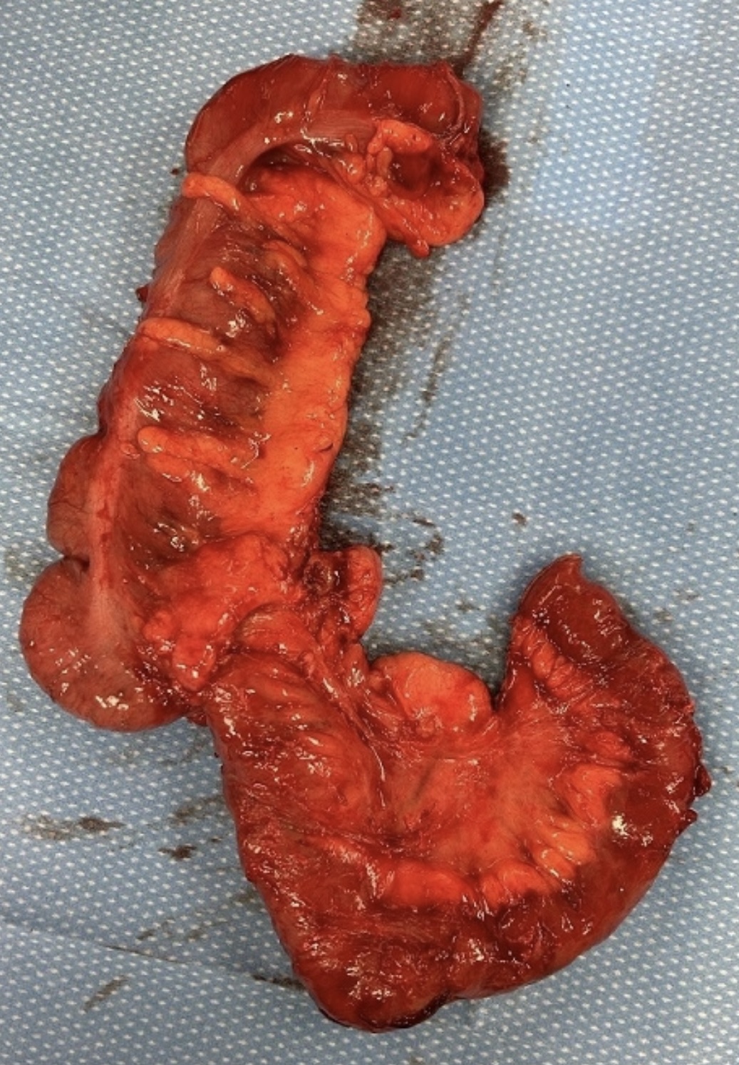
Figure: Gross specimen of right hemi-colectomy consisting of an 18.5 x 6.0 x 5.0 cm cecum with attached 19.0 cm in length by 3.5 cm in diameter terminal ileum. Specimen was opened to reveal a 2.3 x 1.6 x 1.3 cm tan white density on the cecal valve as well as thirty-two lymph nodes measuring up to 1.2 cm in greatest dimension. Final pathology was consistent with benign lymph nodes and extensive small bowel and colonic endometriosis involving the muscularis propria.
Disclosures:
Puja Sasankan indicated no relevant financial relationships.
Vinay Rao indicated no relevant financial relationships.
Marie Borum: Takeda Pharmaceuticals – Consultant, Speakers Bureau.
Samuel Schueler indicated no relevant financial relationships.
Natasha Kamal: Bristol-Myers Squibb – Speakers Bureau. Johnson & Johnson – Consultant. Takeda – Speakers Bureau.
Puja Sasankan, BS1, Vinay Rao, MD1, Marie L.. Borum, MD, MPH, FACG1, Samuel A.. Schueler, MD1, Natasha Kamal, MD2. P4435 - Endometriosis Implantation Masquerading as Stricturing Crohn’s Disease of the Terminal Ileum: A Case Report, ACG 2024 Annual Scientific Meeting Abstracts. Philadelphia, PA: American College of Gastroenterology.
