Tuesday Poster Session
Category: GI Bleeding
P4255 - More Than Meets the Ileum: Adult Melena and Meckel's
Tuesday, October 29, 2024
10:30 AM - 4:00 PM ET
Location: Exhibit Hall E

Has Audio

Ahmad Sandouka
University of Nicosia St. George's Medical School
Nicosia, Nicosia, Cyprus
Presenting Author(s)
Mohammed W Khaled, MD1, Sayed T. Shah, 2, Ahmad Sandouka, 3, Barakat Aburajab Altamimi, MD4
1King Hussein Medical Center, Amman, 'Amman, Jordan; 2University of Nicosia Medical School, Abbotsford, BC, Canada; 3University of Nicosia St. George’s Medical School, Nicosia, Nicosia, Cyprus; 4Mercy Medical Center, Cedar Rapids, IA
Introduction: Acute upper gastrointestinal (GI) bleeding is a prevalent medical emergency associated with significant morbidity and mortality. Peptic ulcer disease represents the most frequent cause of upper GI bleeding. Other potential etiologies include esophagitis, gastritis, esophageal varices, Mallory-Weiss tears, and malignancies, often associated with risk factors like nonsteroidal anti-inflammatory drug (NSAID) use and H. pylori infection.
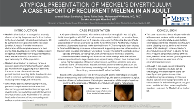
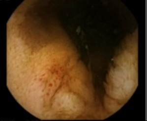
Case Description/Methods: A 42-year-old male presented with melena. Admission hemoglobin was 11.2 g/dL. Initial investigations with esophagogastroduodenoscopy and colonoscopy revealed blood in the terminal ileum, suggesting a small bowel source. A capsule endoscopy the following day identified a polypoid lesion with an ulcerated tip at the 4:36:10 time mark. Additionally, multiple aphthous ulcers were observed in the terminal ileum. A CT enterography scan showed no focal wall thickening or mucosal enhancement, suggesting no active inflammation. A Meckel’s scan was also interestingly unyielding. Notably, the patient reported a similar episode of melena 10 years prior, with an inconclusive workup at that time.
Given the recurrent bleeding and inconclusive initial workup, a double balloon enteroscopy visualized a large diverticulum approximately 100 cm from the ileocecal valve, highly suggestive of Meckel’s diverticulum. Aphthous erosions were also observed in the terminal ileum. Biopsies were obtained from the diverticulum and terminal ileum. Diverticular biopsy revealed gastric heterotopia, while the terminal ileum biopsy demonstrated focal active ileitis.
Based on the visualization of the diverticulum with gastric heterotopia on double balloon enteroscopy and confirmatory biopsy findings, the patient underwent surgical resection of Meckel’s diverticulum. Pathological examination of the surgical specimen confirmed the diagnosis of Meckel’s diverticulum with gastric heterotopia.
Discussion: This case report describes a 42-year-old male with recurrent melena. Initial workup was challenging, but ultimately, double balloon enteroscopy identified Meckel’s diverticulum as the bleeding source. Meckel’s diverticulum, while a well-known cause of GI bleeding in children, is uncommon in adults. Although it can cause classic GI bleeding symptoms, diagnosis in adults can be challenging. In this case, double balloon enteroscopy proved to be a valuable tool for definitive diagnosis. Surgical resection is the standard treatment for Meckel’s diverticulum.

Disclosures:
Mohammed W Khaled, MD1, Sayed T. Shah, 2, Ahmad Sandouka, 3, Barakat Aburajab Altamimi, MD4. P4255 - More Than Meets the Ileum: Adult Melena and Meckel's, ACG 2024 Annual Scientific Meeting Abstracts. Philadelphia, PA: American College of Gastroenterology.
1King Hussein Medical Center, Amman, 'Amman, Jordan; 2University of Nicosia Medical School, Abbotsford, BC, Canada; 3University of Nicosia St. George’s Medical School, Nicosia, Nicosia, Cyprus; 4Mercy Medical Center, Cedar Rapids, IA
Introduction: Acute upper gastrointestinal (GI) bleeding is a prevalent medical emergency associated with significant morbidity and mortality. Peptic ulcer disease represents the most frequent cause of upper GI bleeding. Other potential etiologies include esophagitis, gastritis, esophageal varices, Mallory-Weiss tears, and malignancies, often associated with risk factors like nonsteroidal anti-inflammatory drug (NSAID) use and H. pylori infection.
Case Description/Methods: A 42-year-old male presented with melena. Admission hemoglobin was 11.2 g/dL. Initial investigations with esophagogastroduodenoscopy and colonoscopy revealed blood in the terminal ileum, suggesting a small bowel source. A capsule endoscopy the following day identified a polypoid lesion with an ulcerated tip at the 4:36:10 time mark. Additionally, multiple aphthous ulcers were observed in the terminal ileum. A CT enterography scan showed no focal wall thickening or mucosal enhancement, suggesting no active inflammation. A Meckel’s scan was also interestingly unyielding. Notably, the patient reported a similar episode of melena 10 years prior, with an inconclusive workup at that time.
Given the recurrent bleeding and inconclusive initial workup, a double balloon enteroscopy visualized a large diverticulum approximately 100 cm from the ileocecal valve, highly suggestive of Meckel’s diverticulum. Aphthous erosions were also observed in the terminal ileum. Biopsies were obtained from the diverticulum and terminal ileum. Diverticular biopsy revealed gastric heterotopia, while the terminal ileum biopsy demonstrated focal active ileitis.
Based on the visualization of the diverticulum with gastric heterotopia on double balloon enteroscopy and confirmatory biopsy findings, the patient underwent surgical resection of Meckel’s diverticulum. Pathological examination of the surgical specimen confirmed the diagnosis of Meckel’s diverticulum with gastric heterotopia.
Discussion: This case report describes a 42-year-old male with recurrent melena. Initial workup was challenging, but ultimately, double balloon enteroscopy identified Meckel’s diverticulum as the bleeding source. Meckel’s diverticulum, while a well-known cause of GI bleeding in children, is uncommon in adults. Although it can cause classic GI bleeding symptoms, diagnosis in adults can be challenging. In this case, double balloon enteroscopy proved to be a valuable tool for definitive diagnosis. Surgical resection is the standard treatment for Meckel’s diverticulum.

Figure: Capsule endoscopy showing ulceration.
Disclosures:
Mohammed W Khaled indicated no relevant financial relationships.
Sayed Shah indicated no relevant financial relationships.
Ahmad Sandouka indicated no relevant financial relationships.
Barakat Aburajab Altamimi indicated no relevant financial relationships.
Mohammed W Khaled, MD1, Sayed T. Shah, 2, Ahmad Sandouka, 3, Barakat Aburajab Altamimi, MD4. P4255 - More Than Meets the Ileum: Adult Melena and Meckel's, ACG 2024 Annual Scientific Meeting Abstracts. Philadelphia, PA: American College of Gastroenterology.
