Monday Poster Session
Category: Esophagus
P2244 - Esophagitis in a Post-Liver Transplant Patient: A Case of Cytomegalovirus and Herpes Simplex Virus-1 Coinfection
Monday, October 28, 2024
10:30 AM - 4:00 PM ET
Location: Exhibit Hall E

Has Audio
- AA
Amira Al-Nabolsi, DO
Corewell Health Farmington Hills
Dearborn, MI
Presenting Author(s)
Amira Al-Nabolsi, DO1, Ammad Javaid. Chaudhary, MD2, Taher Jamali, MD3, Allyce N. Caines, MD2, Mazen Elatrache, MD4
1Corewell Health Farmington Hills, Dearborn, MI; 2Henry Ford Health, Detroit, MI; 3Henry Ford Hospital, Detroit, MI; 4Henry Ford Health, Dearborn, MI
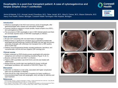
Introduction: Infectious esophagitis is the third most common cause of esophagitis, after gastroesophageal reflux disease and eosinophilic esophagitis. Common infectious organisms include candida, herpes simplex virus (HSV) and cytomegalovirus (CMV). While the occurrence of CMV esophagitis is rare in CMV infected patients, there remains minimal reported cases regarding CMV/HSV esophagitis co-infection. We present a rare case of CMV/HSV esophagitis successfully treated with antivirals.
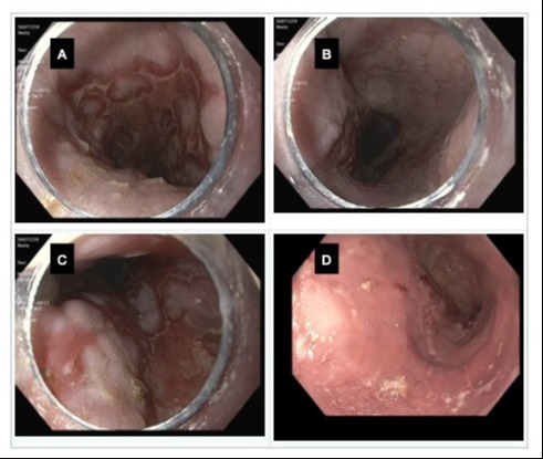
Case Description/Methods: A 59 year old female with history of primary sclerosing cholangitis, who underwent orthotopic liver transplantation from a CMV seropositive donor six years ago, and achalasia secondary to scleroderma, treated with esophageal perusal endoscopic myotomy (EPOEM) one year ago, presented with a one-week history of dysphagia. The patient’s immunosuppressive therapy at the time of presentation included prednisone, tacrolimus, and mycophenolate. The patient had negative CMV serology prior. An esophagogastroduodenoscopy (EGD) revealed severe esophagitis with extensive serpiginous and confluent non-bleeding ulceration (Figure 1A, B, C). Biopsies from the ulcer bed as well as ulcer edges confirmed CMV and herpes simplex virus-1 (HSV-1) co-infection. CMV quantitation in the blood was 9,523 IU/mL, subsequently becoming undetectable following treatment with valganciclovir. Mycophenolate was temporarily discontinued during treatment for CMV and HSV esophagitis. A repeat EGD performed two months later showed esophageal ulcers with no recent bleeding stigmata (Figure 1D). Biopsies revealed candida esophagitis with ulceration, and notably, CMV/HSV testing was negative.
Discussion: CMV/HSV co-infection in the esophagus is very rare and can be associated with higher complication rates including perforation and bleeding. Thus, in immunocompromised hosts with esophagitis, a high index of suspicion for these conditions can help with targeting of appropriate biopsies of the esophagus to yield accurate and early diagnoses, allowing for rapid treatment.

Disclosures:
Amira Al-Nabolsi, DO1, Ammad Javaid. Chaudhary, MD2, Taher Jamali, MD3, Allyce N. Caines, MD2, Mazen Elatrache, MD4. P2244 - Esophagitis in a Post-Liver Transplant Patient: A Case of Cytomegalovirus and Herpes Simplex Virus-1 Coinfection, ACG 2024 Annual Scientific Meeting Abstracts. Philadelphia, PA: American College of Gastroenterology.
1Corewell Health Farmington Hills, Dearborn, MI; 2Henry Ford Health, Detroit, MI; 3Henry Ford Hospital, Detroit, MI; 4Henry Ford Health, Dearborn, MI
Introduction: Infectious esophagitis is the third most common cause of esophagitis, after gastroesophageal reflux disease and eosinophilic esophagitis. Common infectious organisms include candida, herpes simplex virus (HSV) and cytomegalovirus (CMV). While the occurrence of CMV esophagitis is rare in CMV infected patients, there remains minimal reported cases regarding CMV/HSV esophagitis co-infection. We present a rare case of CMV/HSV esophagitis successfully treated with antivirals.
Case Description/Methods: A 59 year old female with history of primary sclerosing cholangitis, who underwent orthotopic liver transplantation from a CMV seropositive donor six years ago, and achalasia secondary to scleroderma, treated with esophageal perusal endoscopic myotomy (EPOEM) one year ago, presented with a one-week history of dysphagia. The patient’s immunosuppressive therapy at the time of presentation included prednisone, tacrolimus, and mycophenolate. The patient had negative CMV serology prior. An esophagogastroduodenoscopy (EGD) revealed severe esophagitis with extensive serpiginous and confluent non-bleeding ulceration (Figure 1A, B, C). Biopsies from the ulcer bed as well as ulcer edges confirmed CMV and herpes simplex virus-1 (HSV-1) co-infection. CMV quantitation in the blood was 9,523 IU/mL, subsequently becoming undetectable following treatment with valganciclovir. Mycophenolate was temporarily discontinued during treatment for CMV and HSV esophagitis. A repeat EGD performed two months later showed esophageal ulcers with no recent bleeding stigmata (Figure 1D). Biopsies revealed candida esophagitis with ulceration, and notably, CMV/HSV testing was negative.
Discussion: CMV/HSV co-infection in the esophagus is very rare and can be associated with higher complication rates including perforation and bleeding. Thus, in immunocompromised hosts with esophagitis, a high index of suspicion for these conditions can help with targeting of appropriate biopsies of the esophagus to yield accurate and early diagnoses, allowing for rapid treatment.

Figure: A: middle third of the esophagus B: Lower third of the esophagus C: Lower third of the esophagus D: Candida esophagitis (lower third of the esophagus).
Disclosures:
Amira Al-Nabolsi indicated no relevant financial relationships.
Ammad Chaudhary indicated no relevant financial relationships.
Taher Jamali indicated no relevant financial relationships.
Allyce Caines indicated no relevant financial relationships.
Mazen Elatrache indicated no relevant financial relationships.
Amira Al-Nabolsi, DO1, Ammad Javaid. Chaudhary, MD2, Taher Jamali, MD3, Allyce N. Caines, MD2, Mazen Elatrache, MD4. P2244 - Esophagitis in a Post-Liver Transplant Patient: A Case of Cytomegalovirus and Herpes Simplex Virus-1 Coinfection, ACG 2024 Annual Scientific Meeting Abstracts. Philadelphia, PA: American College of Gastroenterology.
