Monday Poster Session
Category: Colon
P2085 - True or False? A Case of Large Bowel Obstruction Secondary to Right-Sided Colonic Malignancy Initially Presenting as Ogilvie Syndrome
Monday, October 28, 2024
10:30 AM - 4:00 PM ET
Location: Exhibit Hall E

Has Audio

Brandon Stahl, MD
Albany Medical Center
Albany, NY
Presenting Author(s)
Brandon Stahl, MD, Anika Paradkar, MD, Asra Batool, MD
Albany Medical Center, Albany, NY
Introduction: Colorectal cancer accounts for more than 60% of large bowel obstructions (LBO) and can be a life-threatening surgical emergency with a high morbidity and mortality rate. While plain radiographs provide useful information in the initial evaluation for bowel obstruction, computed tomography (CT) is the gold standard tool for LBO with a sensitivity of 96% and specificity of 93%. Our case presents a patient with initial findings concerning for bowel obstruction vs pseudo-obstruction, who was found to have a right-sided colonic malignancy that was undetected over a series of CTs.
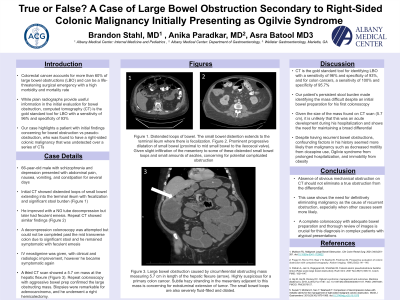
Case Description/Methods: A 66-year-old male with schizophrenia and depression presented with abdominal pain, nausea, vomiting, and constipation for several days. Initial CT showed distended loops of small bowel extending into the terminal ileum with fecalization and significant stool burden (Figure 1). He improved with a NG tube decompression but later had feculent emesis. Repeat CT showed similar findings (Figure 2). A decompression colonoscopy was attempted but could not be completed past the mid transverse colon due to significant stool. He remained symptomatic with feculent emesis. IV neostigmine was given, with clinical and radiologic improvement, however he became symptomatic again. Another CT scan showed a 5.7 cm mass at the hepatic flexure (Figure 3). Repeat colonoscopy with aggressive bowel prep confirmed the large obstructing mass. Biopsies were remarkable for adenocarcinoma, and he underwent a right hemicolectomy.
Discussion: CT is the gold standard tool for identifying LBO with a sensitivity of 96% and specificity of 93%, and for colon cancers, a sensitivity of 100% and specificity of 95.7%. Our patient had four CT scans over two weeks. The first 3 had poor bowel prep and did not find a mass. The last was done with a complete bowel prep and found the 5.7cm mass. Despite having recurrent bowel obstructions, confounding factors in his history seemed more likely than malignancy such as decreased motility from clozapine use, Ogilvie syndrome from prolonged hospitalization, and immobility from obesity. Colon cancer is the most common cause of LBO and was the source of our patient’s bowel obstruction. This case shows the need for definitively eliminating malignancy as the cause of recurrent obstruction, especially when other causes seem more likely. A complete colonoscopy with adequate bowel preparation and thorough review of images is crucial for this diagnosis in complex patients with atypical presentations.

Disclosures:
Brandon Stahl, MD, Anika Paradkar, MD, Asra Batool, MD. P2085 - True or False? A Case of Large Bowel Obstruction Secondary to Right-Sided Colonic Malignancy Initially Presenting as Ogilvie Syndrome, ACG 2024 Annual Scientific Meeting Abstracts. Philadelphia, PA: American College of Gastroenterology.
Albany Medical Center, Albany, NY
Introduction: Colorectal cancer accounts for more than 60% of large bowel obstructions (LBO) and can be a life-threatening surgical emergency with a high morbidity and mortality rate. While plain radiographs provide useful information in the initial evaluation for bowel obstruction, computed tomography (CT) is the gold standard tool for LBO with a sensitivity of 96% and specificity of 93%. Our case presents a patient with initial findings concerning for bowel obstruction vs pseudo-obstruction, who was found to have a right-sided colonic malignancy that was undetected over a series of CTs.
Case Description/Methods: A 66-year-old male with schizophrenia and depression presented with abdominal pain, nausea, vomiting, and constipation for several days. Initial CT showed distended loops of small bowel extending into the terminal ileum with fecalization and significant stool burden (Figure 1). He improved with a NG tube decompression but later had feculent emesis. Repeat CT showed similar findings (Figure 2). A decompression colonoscopy was attempted but could not be completed past the mid transverse colon due to significant stool. He remained symptomatic with feculent emesis. IV neostigmine was given, with clinical and radiologic improvement, however he became symptomatic again. Another CT scan showed a 5.7 cm mass at the hepatic flexure (Figure 3). Repeat colonoscopy with aggressive bowel prep confirmed the large obstructing mass. Biopsies were remarkable for adenocarcinoma, and he underwent a right hemicolectomy.
Discussion: CT is the gold standard tool for identifying LBO with a sensitivity of 96% and specificity of 93%, and for colon cancers, a sensitivity of 100% and specificity of 95.7%. Our patient had four CT scans over two weeks. The first 3 had poor bowel prep and did not find a mass. The last was done with a complete bowel prep and found the 5.7cm mass. Despite having recurrent bowel obstructions, confounding factors in his history seemed more likely than malignancy such as decreased motility from clozapine use, Ogilvie syndrome from prolonged hospitalization, and immobility from obesity. Colon cancer is the most common cause of LBO and was the source of our patient’s bowel obstruction. This case shows the need for definitively eliminating malignancy as the cause of recurrent obstruction, especially when other causes seem more likely. A complete colonoscopy with adequate bowel preparation and thorough review of images is crucial for this diagnosis in complex patients with atypical presentations.

Figure: Figure 1. Distended loops of bowel. The small bowel distention extends to the terminal ileum where there is fecalization. Figure 2. Prominent progressive dilatation of small bowel (proximal to mid small bowel to the ileocecal valve). Given slight infiltration of the mesentery to some of these distended small bowel loops and small amounts of ascites, concerning for potential complicated obstruction. Figure 3. Large bowel obstruction caused by circumferential obstructing mass measuring 5.7 cm in length of the hepatic flexure (arrow). Highly suspicious for a primary colon cancer. Subtle hazy stranding in the mesentery adjacent to this mass is concerning for extraluminal extension of tumor. The small bowel loops are also severely fluid-filled and dilated.
Disclosures:
Brandon Stahl indicated no relevant financial relationships.
Anika Paradkar indicated no relevant financial relationships.
Asra Batool indicated no relevant financial relationships.
Brandon Stahl, MD, Anika Paradkar, MD, Asra Batool, MD. P2085 - True or False? A Case of Large Bowel Obstruction Secondary to Right-Sided Colonic Malignancy Initially Presenting as Ogilvie Syndrome, ACG 2024 Annual Scientific Meeting Abstracts. Philadelphia, PA: American College of Gastroenterology.
