Sunday Poster Session
Category: Biliary/Pancreas
P0132 - Subvesical Bile Duct Leak Post-Cholecystectomy: The Ducts of Luschka
Sunday, October 27, 2024
3:30 PM - 7:00 PM ET
Location: Exhibit Hall E

Has Audio
- PG
Priyatham Gurram, MD
Central Michigan University College of Medicine
Saginaw, MI
Presenting Author(s)
Priyatham Gurram, MD1, Terry Jue, MD2
1Central Michigan University College of Medicine, Saginaw, MI; 2Mayo Clinic, Scottsdale, AZ
Introduction: Bile leaks are an uncommon complication post-cholecystectomy, typically originating from the cystic duct remnant. We present a case of a subvesical bile duct leak after cholecystectomy.
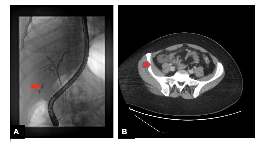
Case Description/Methods: A 40-year-old female developed severe right upper quadrant pain radiating to her right shoulder, accompanied by abdominal distention and bloating, nine days post-laparoscopic cholecystectomy. Initial investigations were unremarkable, and she was discharged with analgesics. She returned to the hospital with worsening pain, chills, and vomiting. A CT scan with oral contrast revealed increased fluid collection in the pelvis and right lower paracolic gutter, suggesting a biliary leak. Her WBC count had risen to 24,000, and liver function tests were abnormal. She underwent IR-guided drainage, which showed reddish-yellow fluid indicating biliary content. A subsequent HIDA scan showed tracer in the right paracolic gutter, confirming a bile leak. Upon transfer to our facility, the patient was started on broad-spectrum antibiotics and ERCP requested. During ERCP, the pancreatic duct was first inadvertently wire cannulated. A second wire was therefore used to access the common bile duct, and cholangiogram showed a leak from small peripheral bile ducts arising from the right posterior hepatic duct. Biliary sphincterotomy was performed, then placement of a plastic biliary stent (10 F x 10 cm) in the common bile duct. A pancreatic duct stent (5 F x 3 cm) was placed as prophylaxis against post-ERCP pancreatitis.
Discussion: Bile leaks post-cholecystectomy most commonly occur at the cystic duct remnant. Much less commonly will the leak occur from small subvesical bile ducts (i.e., the “ducts of Luschka”) communicating between the liver and gallbladder. Imaging studies, particularly the HIDA scan and ERCP, were crucial in confirming the source of the leak. HIDA scan is effective in demonstrating active bile leaks. ERCP is critical in confirming the site of the leak and facilitating therapeutic intervention through biliary sphincterotomy and stent placement. Good fluoroscopic imaging and performance of an excellent cholangiogram is essential in making a diagnosis. Management of bile leaks involves drainage, antibiotics, and ERCP. The leak stopped after stent placement, with plan for stent removal after 2 to 3 months.

Disclosures:
Priyatham Gurram, MD1, Terry Jue, MD2. P0132 - Subvesical Bile Duct Leak Post-Cholecystectomy: The Ducts of Luschka, ACG 2024 Annual Scientific Meeting Abstracts. Philadelphia, PA: American College of Gastroenterology.
1Central Michigan University College of Medicine, Saginaw, MI; 2Mayo Clinic, Scottsdale, AZ
Introduction: Bile leaks are an uncommon complication post-cholecystectomy, typically originating from the cystic duct remnant. We present a case of a subvesical bile duct leak after cholecystectomy.
Case Description/Methods: A 40-year-old female developed severe right upper quadrant pain radiating to her right shoulder, accompanied by abdominal distention and bloating, nine days post-laparoscopic cholecystectomy. Initial investigations were unremarkable, and she was discharged with analgesics. She returned to the hospital with worsening pain, chills, and vomiting. A CT scan with oral contrast revealed increased fluid collection in the pelvis and right lower paracolic gutter, suggesting a biliary leak. Her WBC count had risen to 24,000, and liver function tests were abnormal. She underwent IR-guided drainage, which showed reddish-yellow fluid indicating biliary content. A subsequent HIDA scan showed tracer in the right paracolic gutter, confirming a bile leak. Upon transfer to our facility, the patient was started on broad-spectrum antibiotics and ERCP requested. During ERCP, the pancreatic duct was first inadvertently wire cannulated. A second wire was therefore used to access the common bile duct, and cholangiogram showed a leak from small peripheral bile ducts arising from the right posterior hepatic duct. Biliary sphincterotomy was performed, then placement of a plastic biliary stent (10 F x 10 cm) in the common bile duct. A pancreatic duct stent (5 F x 3 cm) was placed as prophylaxis against post-ERCP pancreatitis.
Discussion: Bile leaks post-cholecystectomy most commonly occur at the cystic duct remnant. Much less commonly will the leak occur from small subvesical bile ducts (i.e., the “ducts of Luschka”) communicating between the liver and gallbladder. Imaging studies, particularly the HIDA scan and ERCP, were crucial in confirming the source of the leak. HIDA scan is effective in demonstrating active bile leaks. ERCP is critical in confirming the site of the leak and facilitating therapeutic intervention through biliary sphincterotomy and stent placement. Good fluoroscopic imaging and performance of an excellent cholangiogram is essential in making a diagnosis. Management of bile leaks involves drainage, antibiotics, and ERCP. The leak stopped after stent placement, with plan for stent removal after 2 to 3 months.

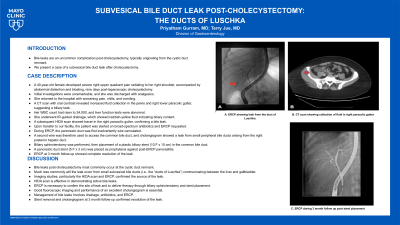
Figure: A. ERCP showing leak from the duct of Luschka. B. CT scan showing collection of fluid in right paracolic gutter.
Disclosures:
Priyatham Gurram indicated no relevant financial relationships.
Terry Jue: Boston Scientific – Instructor.
Priyatham Gurram, MD1, Terry Jue, MD2. P0132 - Subvesical Bile Duct Leak Post-Cholecystectomy: The Ducts of Luschka, ACG 2024 Annual Scientific Meeting Abstracts. Philadelphia, PA: American College of Gastroenterology.
