Sunday Poster Session
Category: Biliary/Pancreas
P0143 - It's Not a Tumor! Unveiling Xanthogranulomatous Cholecystitis in the Quest for Cancer Diagnosis
Sunday, October 27, 2024
3:30 PM - 7:00 PM ET
Location: Exhibit Hall E

Has Audio
- PS
Pranay Siriya, MD
Maimonides Medical Center
Brooklyn, NY
Presenting Author(s)
Pranay Siriya, MD, Diana Cheung, DO, Neha Sharma, MBBS, MD, Ahmed E. Salem, MBBCh, Syed Mujtaba Baqir, MD, Meredith E. Pittman, MD, Yuriy Tsirlin, MD
Maimonides Medical Center, Brooklyn, NY
Introduction: Xanthogranulomatous cholecystitis is a rare inflammatory disease of the gallbladder which on initial presentation can be easily confused with gallbladder carcinoma. We present a case of a patient that was being worked up for a suspected gastrointestinal malignancy and presented with acute cholangitis.
Case Description/Methods: A 79-year-old man with history of hypertension was in the process of being evaluated on an outpatient basis for jaundice and vague right-sided abdominal discomfort ongoing for several months. New onset of fever and chills prompted referral to the emergency room. On initial evaluation, the patient was septic and had liver enzyme pattern consistent with obstructive cholangiopathy: total/direct bilirubin 19.4/14 mg/dL, AST 48, ALT 47, ALP 387.
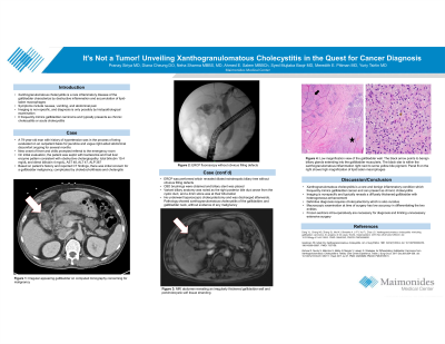
Based on presentation and reported CT findings, initial impression was suspicious for gallbladder cancer, possibly complicated by choledocholithiasis and cholangitis. After stabilization, an ERCP was performed. Dilated extrahepatic biliary tree was noted without obvious filling defects. CBD brushings were obtained and biliary stent was placed. Brushings were unrevealing. Liver enzymes quickly improved.
A subsequent MRI revealed diffuse mildly irregular gallbladder wall thickening, cholelithiasis with mild pericholecystic soft tissue stranding. Variant biliary anatomy was noted as the right posterior bile duct arose from the cystic duct, and a 4mm stone was at their bifurcation.
The patient underwent laparoscopic cholecystectomy and was discharged afterwards. Pathology showed xanthogranulomatous cholecystitis of the gallbladder, and gallbladder neck, without evidence of any malignancy.
Discussion: Xanthogranulomatous cholecystitis is a rare and benign inflammatory condition which frequently mimics gallbladder cancer and can present as chronic cholecystitis. Imaging is nonspecific and typically reveals a diffusely thickened gallbladder with heterogeneous enhancement. Definitive diagnosis requires cholecystectomy which is also curative. Macroscopic examination at time of surgery has low accuracy in differentiating the two entities, and frozen sections intra-operatively are necessary for diagnosis and limiting unnecessary extensive surgery. Final diagnosis requires pathological examination, and if gallbladder cancer is identified, radical cholecystectomy may be necessary.
This case presents a well described, but rare, and therefore easily missed benign entity mimicking a gallbladder cancer, easily treated when recognized correctly.

Disclosures:
Pranay Siriya, MD, Diana Cheung, DO, Neha Sharma, MBBS, MD, Ahmed E. Salem, MBBCh, Syed Mujtaba Baqir, MD, Meredith E. Pittman, MD, Yuriy Tsirlin, MD. P0143 - It's Not a Tumor! Unveiling Xanthogranulomatous Cholecystitis in the Quest for Cancer Diagnosis, ACG 2024 Annual Scientific Meeting Abstracts. Philadelphia, PA: American College of Gastroenterology.
Maimonides Medical Center, Brooklyn, NY
Introduction: Xanthogranulomatous cholecystitis is a rare inflammatory disease of the gallbladder which on initial presentation can be easily confused with gallbladder carcinoma. We present a case of a patient that was being worked up for a suspected gastrointestinal malignancy and presented with acute cholangitis.
Case Description/Methods: A 79-year-old man with history of hypertension was in the process of being evaluated on an outpatient basis for jaundice and vague right-sided abdominal discomfort ongoing for several months. New onset of fever and chills prompted referral to the emergency room. On initial evaluation, the patient was septic and had liver enzyme pattern consistent with obstructive cholangiopathy: total/direct bilirubin 19.4/14 mg/dL, AST 48, ALT 47, ALP 387.
Based on presentation and reported CT findings, initial impression was suspicious for gallbladder cancer, possibly complicated by choledocholithiasis and cholangitis. After stabilization, an ERCP was performed. Dilated extrahepatic biliary tree was noted without obvious filling defects. CBD brushings were obtained and biliary stent was placed. Brushings were unrevealing. Liver enzymes quickly improved.
A subsequent MRI revealed diffuse mildly irregular gallbladder wall thickening, cholelithiasis with mild pericholecystic soft tissue stranding. Variant biliary anatomy was noted as the right posterior bile duct arose from the cystic duct, and a 4mm stone was at their bifurcation.
The patient underwent laparoscopic cholecystectomy and was discharged afterwards. Pathology showed xanthogranulomatous cholecystitis of the gallbladder, and gallbladder neck, without evidence of any malignancy.
Discussion: Xanthogranulomatous cholecystitis is a rare and benign inflammatory condition which frequently mimics gallbladder cancer and can present as chronic cholecystitis. Imaging is nonspecific and typically reveals a diffusely thickened gallbladder with heterogeneous enhancement. Definitive diagnosis requires cholecystectomy which is also curative. Macroscopic examination at time of surgery has low accuracy in differentiating the two entities, and frozen sections intra-operatively are necessary for diagnosis and limiting unnecessary extensive surgery. Final diagnosis requires pathological examination, and if gallbladder cancer is identified, radical cholecystectomy may be necessary.
This case presents a well described, but rare, and therefore easily missed benign entity mimicking a gallbladder cancer, easily treated when recognized correctly.

Figure: Panel A is a low magnification view of the gallbladder wall. The black arrow points to benign biliary glands extending into the gallbladder muscularis. The black star is within the xanthogranulomatous inflammation right next to some yellow bile pigment. Panel B shows a high magnification image of the lipid-laden macrophages of the inflammatory mass. Panel C shows an irregular appearing gallbladder on computed tomography concerning for malignancy.
Disclosures:
Pranay Siriya indicated no relevant financial relationships.
Diana Cheung indicated no relevant financial relationships.
Neha Sharma indicated no relevant financial relationships.
Ahmed Salem indicated no relevant financial relationships.
Syed Mujtaba Baqir indicated no relevant financial relationships.
Meredith Pittman indicated no relevant financial relationships.
Yuriy Tsirlin indicated no relevant financial relationships.
Pranay Siriya, MD, Diana Cheung, DO, Neha Sharma, MBBS, MD, Ahmed E. Salem, MBBCh, Syed Mujtaba Baqir, MD, Meredith E. Pittman, MD, Yuriy Tsirlin, MD. P0143 - It's Not a Tumor! Unveiling Xanthogranulomatous Cholecystitis in the Quest for Cancer Diagnosis, ACG 2024 Annual Scientific Meeting Abstracts. Philadelphia, PA: American College of Gastroenterology.
