Sunday Poster Session
Category: Biliary/Pancreas
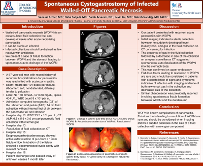
P0173 - Spontaneous Cystogastrostomy of Infected Walled-Off Pancreatic Necrosis
Sunday, October 27, 2024
3:30 PM - 7:00 PM ET
Location: Exhibit Hall E

Has Audio

Vanessa F. Eller, MD
University of Arizona College of Medicine
Phoenix, AZ
Presenting Author(s)
Vanessa F. Eller, MD1, Raha Sadjadi, MD2, Sarah Arvaneh, DO3, Kevin Liu, MD1, Rakesh Nanda, MD, FACG4
1University of Arizona College of Medicine, Phoenix, AZ; 2Banner University Medical Center, Phoenix, AZ; 3Banner - University of Arizona, Phoenix, AZ; 4Carl T. Hayden Veterans' Administration Medical Center, Phoenix, AZ
Introduction: Walled-off pancreatic necrosis (WOPN), a complication of acute pancreatitis usually developing 4 weeks after an initial episode of pancreatitis, is an encapsulated fluid collection that can be either infectious or sterile. Infected collections should be drained as few resolve with antibiotics alone. We describe a case of spontaneous auto-drainage of WOPN after fistula formation between the fluid collection and the stomach.
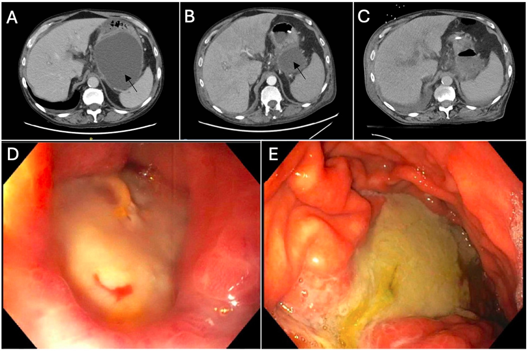
Case Description/Methods: A 57-year-old man with alcohol and tobacco use disorder and recent history of recurrent hospitalizations for acute pancreatitis was readmitted to the hospital with pancreatitis. On presentation, his heart rate was 104 beats per minute and his abdomen was soft and nondistended with diffuse tenderness to palpation. Labs showed sodium 130 mmol/L, creatinine 0.68 mg/dL, lipase 404 U/L, and white blood cell (WBC) count 8 x 10³ per microliter (uL). Computed tomography (CT) of the abdomen and pelvis (A&P) with intravenous (IV) contrast showed a 14 cm organized fluid collection without internal foci of air between the pancreatic tail and stomach. Ten days later, his WBC acutely rose to 23.5 x 10³ per uL. CT A&P showed a now 6.5 x 5.9 x 3.0 cm peripancreatic fluid collection with new gas. The next day he was febrile and bacteremic, so he was started on IV antibiotics. Repeat abdominal CT showed no pancreatic fluid collection. An esophagogastroduodenoscopy showed a fistula in the proximal greater curvature of the stomach with active extravasation of pus from a 12mm fistula orifice. Intubation of the fistula showed a decompressed cystic cavity with minimal necrosis. The patient was discharged on hospital day 18. He passed away 1 month later.
Discussion: In our patient with WOPN, initial abdominal imaging indicated a sterile fluid collection. However, the acute onset of fever, leukocytosis, gas in the fluid collection, and bacteremia was highly suggestive of an infectious etiology. While our attention was diverted to the gas in the fluid collection, it was suspicious that repeat CT scan showed a smaller fluid collection. In hindsight, this smaller collection with gas was indicative of auto-fistulization of the WOPN into the adjacent stomach body. A similar phenomenon was previously reported involving spontaneous fistula formation between WOPN and the duodenum (Boopathy et al. 2022). Fistulous tracts leading to resolution of WOPN are rare and should be considered when imaging shows a sudden decrease in the size of a fluid collection with a new gas component.

Disclosures:
Vanessa F. Eller, MD1, Raha Sadjadi, MD2, Sarah Arvaneh, DO3, Kevin Liu, MD1, Rakesh Nanda, MD, FACG4. P0173 - Spontaneous Cystogastrostomy of Infected Walled-Off Pancreatic Necrosis, ACG 2024 Annual Scientific Meeting Abstracts. Philadelphia, PA: American College of Gastroenterology.
1University of Arizona College of Medicine, Phoenix, AZ; 2Banner University Medical Center, Phoenix, AZ; 3Banner - University of Arizona, Phoenix, AZ; 4Carl T. Hayden Veterans' Administration Medical Center, Phoenix, AZ
Introduction: Walled-off pancreatic necrosis (WOPN), a complication of acute pancreatitis usually developing 4 weeks after an initial episode of pancreatitis, is an encapsulated fluid collection that can be either infectious or sterile. Infected collections should be drained as few resolve with antibiotics alone. We describe a case of spontaneous auto-drainage of WOPN after fistula formation between the fluid collection and the stomach.
Case Description/Methods: A 57-year-old man with alcohol and tobacco use disorder and recent history of recurrent hospitalizations for acute pancreatitis was readmitted to the hospital with pancreatitis. On presentation, his heart rate was 104 beats per minute and his abdomen was soft and nondistended with diffuse tenderness to palpation. Labs showed sodium 130 mmol/L, creatinine 0.68 mg/dL, lipase 404 U/L, and white blood cell (WBC) count 8 x 10³ per microliter (uL). Computed tomography (CT) of the abdomen and pelvis (A&P) with intravenous (IV) contrast showed a 14 cm organized fluid collection without internal foci of air between the pancreatic tail and stomach. Ten days later, his WBC acutely rose to 23.5 x 10³ per uL. CT A&P showed a now 6.5 x 5.9 x 3.0 cm peripancreatic fluid collection with new gas. The next day he was febrile and bacteremic, so he was started on IV antibiotics. Repeat abdominal CT showed no pancreatic fluid collection. An esophagogastroduodenoscopy showed a fistula in the proximal greater curvature of the stomach with active extravasation of pus from a 12mm fistula orifice. Intubation of the fistula showed a decompressed cystic cavity with minimal necrosis. The patient was discharged on hospital day 18. He passed away 1 month later.
Discussion: In our patient with WOPN, initial abdominal imaging indicated a sterile fluid collection. However, the acute onset of fever, leukocytosis, gas in the fluid collection, and bacteremia was highly suggestive of an infectious etiology. While our attention was diverted to the gas in the fluid collection, it was suspicious that repeat CT scan showed a smaller fluid collection. In hindsight, this smaller collection with gas was indicative of auto-fistulization of the WOPN into the adjacent stomach body. A similar phenomenon was previously reported involving spontaneous fistula formation between WOPN and the duodenum (Boopathy et al. 2022). Fistulous tracts leading to resolution of WOPN are rare and should be considered when imaging shows a sudden decrease in the size of a fluid collection with a new gas component.

Figure: Figure: Computed tomography of the abdomen and pelvis showing the progression of the walled-off pancreatic necrosis (WOPN): (A) the WOPN as marked by the black arrow, (B) a spontaneous decrease in size of the WOPN, and (C) evidence of WOPN resolution. (D) Endoscopic visualization of the fistula in the proximal greater curvature of the stomach with active extravasation from a 12mm fistula orifice. (E) Endoscopic evidence of drainage in the gastric body which originated from the WOPN.
Disclosures:
Vanessa Eller indicated no relevant financial relationships.
Raha Sadjadi indicated no relevant financial relationships.
Sarah Arvaneh indicated no relevant financial relationships.
Kevin Liu indicated no relevant financial relationships.
Rakesh Nanda indicated no relevant financial relationships.
Vanessa F. Eller, MD1, Raha Sadjadi, MD2, Sarah Arvaneh, DO3, Kevin Liu, MD1, Rakesh Nanda, MD, FACG4. P0173 - Spontaneous Cystogastrostomy of Infected Walled-Off Pancreatic Necrosis, ACG 2024 Annual Scientific Meeting Abstracts. Philadelphia, PA: American College of Gastroenterology.
