Sunday Poster Session
Category: Colon
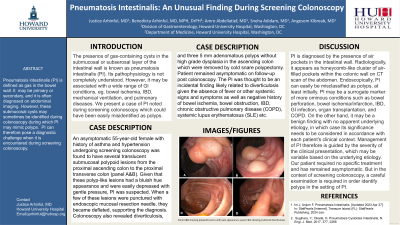
P0250 - Pneumatosis Intestinalis: An Unusual Finding During Screening Colonoscopy
Sunday, October 27, 2024
3:30 PM - 7:00 PM ET
Location: Exhibit Hall E

Has Audio
- JA
Justice Arhinful, MD
Howard University Hospital
Washington, DC
Presenting Author(s)
Justice Arhinful, MD, Benedicta Arhinful, MD, MPH, DrPH, Amro Abdellatief, MD, Sneha Adidam, MD, Angesom Kibreab, MD
Howard University Hospital, Washington, DC
Introduction: Pneumatosis intestinalis (PI) is a condition characterized by the presence of several gas-containing cysts in the submucosal or subserosal layer of the intestinal wall. The pathophysiology is not completely understood. However, it may be associated with a wide range of gastrointestinal conditions, from intestinal ischemia to inflammatory bowel disease, as well as with endoscopic procedures, mechanical ventilation, and pulmonary diseases. We present a case of PI noted during screening colonoscopy which could be easily misidentified as polyps.
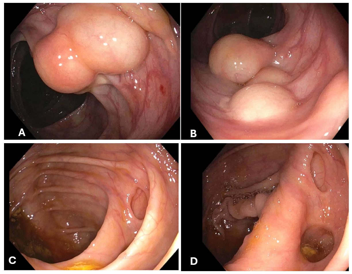
Case Description/Methods: An asymptomatic 55-year-old female with history of asthma and hypertension undergoing screening colonoscopy was found to have several translucent submucosal polypoid lesions from the proximal ascending colon to the proximal transverse colon (panel A&B). Given that these polyp-like lesions had a bluish hue appearance and were easily depressed with gentle pressure, PI was suspected. When a few of these lesions were punctured with endoscopic mucosal resection needle, they became deflated, supporting the diagnosis. Colonoscopy also revealed diverticulosis and three 5 mm adenomatous polyps without high grade dysplasia in the ascending colon which were removed with cold snare polypectomy. Patient remained asymptomatic at his follow-up; the PI was considered to be an incidental finding likely related to diverticulosis given the absence of fever or other systemic signs as well as negative history of bowel ischemia, obstruction, inflammatory bowel disease (IBD), chronic obstructive pulmonary disease (COPD), systemic lupus erythematosus (SLE) etc.
Discussion: PI is diagnosed by the presence of air pockets in the intestinal wall. Radiologically, it appears as honeycomb-like cluster of air-filled pockets within the colonic wall on computed tomographic imaging of the abdomen. Endoscopically, they can easily be misclassified as polyps, at least initially. These air-filled pockets may be a surrogate marker of more ominous medical conditions such as bowel perforation, bowel ischemia/infarction, IBD, GI infection, organ transplantation, and COPD. On the other hand, it may be a benign finding with no apparent underlying etiology, in which case its significance needs to be considered in accordance with each patient's clinical context. Management of PI therefore is guided by the severity of the clinical presentation, which may be variable based on the underlying etiology. Our patient required no specific treatment and has remained asymptomatic.

Disclosures:
Justice Arhinful, MD, Benedicta Arhinful, MD, MPH, DrPH, Amro Abdellatief, MD, Sneha Adidam, MD, Angesom Kibreab, MD. P0250 - Pneumatosis Intestinalis: An Unusual Finding During Screening Colonoscopy, ACG 2024 Annual Scientific Meeting Abstracts. Philadelphia, PA: American College of Gastroenterology.
Howard University Hospital, Washington, DC
Introduction: Pneumatosis intestinalis (PI) is a condition characterized by the presence of several gas-containing cysts in the submucosal or subserosal layer of the intestinal wall. The pathophysiology is not completely understood. However, it may be associated with a wide range of gastrointestinal conditions, from intestinal ischemia to inflammatory bowel disease, as well as with endoscopic procedures, mechanical ventilation, and pulmonary diseases. We present a case of PI noted during screening colonoscopy which could be easily misidentified as polyps.
Case Description/Methods: An asymptomatic 55-year-old female with history of asthma and hypertension undergoing screening colonoscopy was found to have several translucent submucosal polypoid lesions from the proximal ascending colon to the proximal transverse colon (panel A&B). Given that these polyp-like lesions had a bluish hue appearance and were easily depressed with gentle pressure, PI was suspected. When a few of these lesions were punctured with endoscopic mucosal resection needle, they became deflated, supporting the diagnosis. Colonoscopy also revealed diverticulosis and three 5 mm adenomatous polyps without high grade dysplasia in the ascending colon which were removed with cold snare polypectomy. Patient remained asymptomatic at his follow-up; the PI was considered to be an incidental finding likely related to diverticulosis given the absence of fever or other systemic signs as well as negative history of bowel ischemia, obstruction, inflammatory bowel disease (IBD), chronic obstructive pulmonary disease (COPD), systemic lupus erythematosus (SLE) etc.
Discussion: PI is diagnosed by the presence of air pockets in the intestinal wall. Radiologically, it appears as honeycomb-like cluster of air-filled pockets within the colonic wall on computed tomographic imaging of the abdomen. Endoscopically, they can easily be misclassified as polyps, at least initially. These air-filled pockets may be a surrogate marker of more ominous medical conditions such as bowel perforation, bowel ischemia/infarction, IBD, GI infection, organ transplantation, and COPD. On the other hand, it may be a benign finding with no apparent underlying etiology, in which case its significance needs to be considered in accordance with each patient's clinical context. Management of PI therefore is guided by the severity of the clinical presentation, which may be variable based on the underlying etiology. Our patient required no specific treatment and has remained asymptomatic.

Figure: Panel A&B showing polypoid lesions with pale appearance; panel C&D showing scattered diverticulosis
Disclosures:
Justice Arhinful indicated no relevant financial relationships.
Benedicta Arhinful indicated no relevant financial relationships.
Amro Abdellatief indicated no relevant financial relationships.
Sneha Adidam indicated no relevant financial relationships.
Angesom Kibreab indicated no relevant financial relationships.
Justice Arhinful, MD, Benedicta Arhinful, MD, MPH, DrPH, Amro Abdellatief, MD, Sneha Adidam, MD, Angesom Kibreab, MD. P0250 - Pneumatosis Intestinalis: An Unusual Finding During Screening Colonoscopy, ACG 2024 Annual Scientific Meeting Abstracts. Philadelphia, PA: American College of Gastroenterology.

