Sunday Poster Session
Category: Colon
P0253 - Full Thickness Resection of a Rare Cecal Granular Cell Tumor
Sunday, October 27, 2024
3:30 PM - 7:00 PM ET
Location: Exhibit Hall E

Has Audio

Nishith Sagubadi, BS
Wayne State University School of Medicine
Detroit, MI
Presenting Author(s)
Nishith Sagubadi, BS1, Faisal Nimri, MD2, Taher Jamali, MD2, Cyrus Piraka, MD3, Syed-Mohammed Jafri, MD3
1Wayne State University School of Medicine, Detroit, MI; 2Henry Ford Hospital, Detroit, MI; 3Henry Ford Health, Detroit, MI
Introduction: We present an interesting case of a 40 year old patient who underwent colonoscopy and was found to have a granular cell tumor in the cecum.
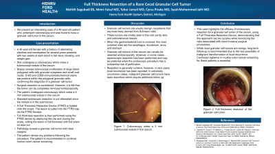
Case Description/Methods: Our patient is a wonderful 40 year old female with a history of alternating diarrhea and constipation for several years. She had two weeks of dark blood in her stool, bloating, and weight gain. She undergoes a colonoscopy which notes a submucosal nodule in the cecum. Biopsy reveals submucosal proliferation of large bland polygonal cells with granular cytoplasm and small oval nuclei. S100 and CD68 immunohistochemical stains are positive within the polygonal granular cells, confirming the diagnosis of a granular cell tumor. Surgical resection is considered. However, it is felt that the tumor can be completely removed endoscopically. She undergoes colonoscopy which notes a 5 mm submucosal nodule in the cecum. Standard endoscopic resection is not attempted since the nodule is in the submucosa. A FTRD (Full Thickness Resection Device) is loaded onto the scope. The lesion is pulled into the FTRD cap via the FTRD forceps. Full thickness resection is then performed using the FTRD device by deploying the clip and closing the snare, cutting the lesion in full thickness with hot snare on auto-cut. The patient tolerates the procedure well. Pathology reveals a granular cell tumor with clear margins. The patient denies any problems following the procedure. The patient is recommended to continue routine colon cancer screening.
Discussion: Granular cell tumors are usually benign neoplasms that are most likely derived from Schwann cells. They are mostly seen in the oral cavity, skin, and subcutaneous tissues. When the gastrointestinal tract is involved, the most common sites are the esophagus, duodenum, anus, and stomach. Granular cell tumors of the cecum can usually be resected endoscopically. However, in some cases, laparoscopic resection has been performed, and may be preferred when the endoscopic procedure has a substantial risk of perforation. Resection is generally curative, however, in rare cases local recurrence has been reported. In extremely uncommon cases, malignant granular cell tumors have been described which require additional follow up.
Disclosures:
Nishith Sagubadi, BS1, Faisal Nimri, MD2, Taher Jamali, MD2, Cyrus Piraka, MD3, Syed-Mohammed Jafri, MD3. P0253 - Full Thickness Resection of a Rare Cecal Granular Cell Tumor, ACG 2024 Annual Scientific Meeting Abstracts. Philadelphia, PA: American College of Gastroenterology.
1Wayne State University School of Medicine, Detroit, MI; 2Henry Ford Hospital, Detroit, MI; 3Henry Ford Health, Detroit, MI
Introduction: We present an interesting case of a 40 year old patient who underwent colonoscopy and was found to have a granular cell tumor in the cecum.
Case Description/Methods: Our patient is a wonderful 40 year old female with a history of alternating diarrhea and constipation for several years. She had two weeks of dark blood in her stool, bloating, and weight gain. She undergoes a colonoscopy which notes a submucosal nodule in the cecum. Biopsy reveals submucosal proliferation of large bland polygonal cells with granular cytoplasm and small oval nuclei. S100 and CD68 immunohistochemical stains are positive within the polygonal granular cells, confirming the diagnosis of a granular cell tumor. Surgical resection is considered. However, it is felt that the tumor can be completely removed endoscopically. She undergoes colonoscopy which notes a 5 mm submucosal nodule in the cecum. Standard endoscopic resection is not attempted since the nodule is in the submucosa. A FTRD (Full Thickness Resection Device) is loaded onto the scope. The lesion is pulled into the FTRD cap via the FTRD forceps. Full thickness resection is then performed using the FTRD device by deploying the clip and closing the snare, cutting the lesion in full thickness with hot snare on auto-cut. The patient tolerates the procedure well. Pathology reveals a granular cell tumor with clear margins. The patient denies any problems following the procedure. The patient is recommended to continue routine colon cancer screening.
Discussion: Granular cell tumors are usually benign neoplasms that are most likely derived from Schwann cells. They are mostly seen in the oral cavity, skin, and subcutaneous tissues. When the gastrointestinal tract is involved, the most common sites are the esophagus, duodenum, anus, and stomach. Granular cell tumors of the cecum can usually be resected endoscopically. However, in some cases, laparoscopic resection has been performed, and may be preferred when the endoscopic procedure has a substantial risk of perforation. Resection is generally curative, however, in rare cases local recurrence has been reported. In extremely uncommon cases, malignant granular cell tumors have been described which require additional follow up.
Disclosures:
Nishith Sagubadi indicated no relevant financial relationships.
Faisal Nimri indicated no relevant financial relationships.
Taher Jamali indicated no relevant financial relationships.
Cyrus Piraka: Aries and US Endoscopy – Grant/Research Support. NIH – Grant/Research Support.
Syed-Mohammed Jafri: Gilead, Takeda, Abbvie, Intercept, VectivBio – Advisor or Review Panel Member, Speakers Bureau.
Nishith Sagubadi, BS1, Faisal Nimri, MD2, Taher Jamali, MD2, Cyrus Piraka, MD3, Syed-Mohammed Jafri, MD3. P0253 - Full Thickness Resection of a Rare Cecal Granular Cell Tumor, ACG 2024 Annual Scientific Meeting Abstracts. Philadelphia, PA: American College of Gastroenterology.
