Sunday Poster Session
Category: Colon
P0292 - Beyond the Obvious: Unmasking Rectal Lipoma Misdiagnosed as Hemorrhoids
Sunday, October 27, 2024
3:30 PM - 7:00 PM ET
Location: Exhibit Hall E

Has Audio

Pratiksha Moliya, MD
Jamaica Hospital Medical Center
Edison, NJ
Presenting Author(s)
Pratiksha Moliya, MD1, Hasan Al-Obaidi, MD2, Avani Patel, MD3
1Jamaica Hospital Medical Center, Edison, NJ; 2Jamaica Hospital Medical Center, Briarwood, NY; 3Jamaica Hospital Medical Center, Queens, NY
Introduction: Lipomas are rare benign tumors, constituting about 5% of all gastrointestinal (GI) tumors, with rectal lipomas even rarer, accounting for 3.4% of colorectal lipomas. We present a case initially misdiagnosed as hemorrhoids, later identified as having a rectal lipoma.
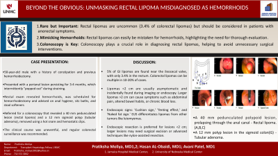
Case Description/Methods: A 50-year-old man with a history of constipation and hemorrhoidectomy presented to the surgical clinic with a perianal lesion for 3-4 months, which occasionally "popped out" while straining. He denied any history of anemia, changes in bowel habits, rectal bleeding, alarming GI symptoms, family history of GI cancer, and having undergone a colonoscopy. Lab results were normal. Rectal examination revealed hemorrhoids. He was scheduled for an examination under anesthesia and hemorrhoidectomy, and was advised on anal hygiene, sitz baths, and stool softeners. Prior to the surgery, he was referred for a colonoscopy that revealed a 40 mm pedunculated lesion (rectal lipoma) and a 12 mm sigmoid polyp (tubular adenoma), removed using a hot snare and hemostatic clips. The diagnosis was confirmed by histology. His clinical course was uneventful. He was advised to undergo regular colorectal surveillance.
Discussion: GI lipomas are rare, primarily found near the ileocecal valve of the ascending colon (45%), with 3.4% located in the rectum. Colorectal lipomas can be multiple in 10-20% of cases. Lipomas under 2 cm are usually asymptomatic, often detected incidentally during imaging or endoscopies as smooth, soft, yellowish masses. Larger lipomas can cause symptoms such as abdominal pain, altered bowel habits, and chronic blood loss, similar to those experienced by our patient, whose symptoms initially mimicked hemorrhoids. The mucosa over a lipoma is usually intact but may show occasional ulcerations and erythema. Endoscopic signs include the 'cushion sign', 'tenting effect', and 'naked fat sign'. EUS differentiates lipomas from leiomyomas. Management typically involves observation for smaller tumors, while larger or symptomatic ones may require resection, ranging from endoscopic removal for lesions under 2 cm to surgical excision for those over 4 cm or causing complications such as intussusception. Endoscopic treatments range from snare resection for smaller tumors to nylon-assisted resection for larger ones. This case underscores the importance of accurate diagnosis in anorectal issues, where symptoms like hemorrhoids can mask rarer conditions like rectal lipomas. Colonoscopy prevented unnecessary surgery in our patient.

Disclosures:
Pratiksha Moliya, MD1, Hasan Al-Obaidi, MD2, Avani Patel, MD3. P0292 - Beyond the Obvious: Unmasking Rectal Lipoma Misdiagnosed as Hemorrhoids, ACG 2024 Annual Scientific Meeting Abstracts. Philadelphia, PA: American College of Gastroenterology.
1Jamaica Hospital Medical Center, Edison, NJ; 2Jamaica Hospital Medical Center, Briarwood, NY; 3Jamaica Hospital Medical Center, Queens, NY
Introduction: Lipomas are rare benign tumors, constituting about 5% of all gastrointestinal (GI) tumors, with rectal lipomas even rarer, accounting for 3.4% of colorectal lipomas. We present a case initially misdiagnosed as hemorrhoids, later identified as having a rectal lipoma.
Case Description/Methods: A 50-year-old man with a history of constipation and hemorrhoidectomy presented to the surgical clinic with a perianal lesion for 3-4 months, which occasionally "popped out" while straining. He denied any history of anemia, changes in bowel habits, rectal bleeding, alarming GI symptoms, family history of GI cancer, and having undergone a colonoscopy. Lab results were normal. Rectal examination revealed hemorrhoids. He was scheduled for an examination under anesthesia and hemorrhoidectomy, and was advised on anal hygiene, sitz baths, and stool softeners. Prior to the surgery, he was referred for a colonoscopy that revealed a 40 mm pedunculated lesion (rectal lipoma) and a 12 mm sigmoid polyp (tubular adenoma), removed using a hot snare and hemostatic clips. The diagnosis was confirmed by histology. His clinical course was uneventful. He was advised to undergo regular colorectal surveillance.
Discussion: GI lipomas are rare, primarily found near the ileocecal valve of the ascending colon (45%), with 3.4% located in the rectum. Colorectal lipomas can be multiple in 10-20% of cases. Lipomas under 2 cm are usually asymptomatic, often detected incidentally during imaging or endoscopies as smooth, soft, yellowish masses. Larger lipomas can cause symptoms such as abdominal pain, altered bowel habits, and chronic blood loss, similar to those experienced by our patient, whose symptoms initially mimicked hemorrhoids. The mucosa over a lipoma is usually intact but may show occasional ulcerations and erythema. Endoscopic signs include the 'cushion sign', 'tenting effect', and 'naked fat sign'. EUS differentiates lipomas from leiomyomas. Management typically involves observation for smaller tumors, while larger or symptomatic ones may require resection, ranging from endoscopic removal for lesions under 2 cm to surgical excision for those over 4 cm or causing complications such as intussusception. Endoscopic treatments range from snare resection for smaller tumors to nylon-assisted resection for larger ones. This case underscores the importance of accurate diagnosis in anorectal issues, where symptoms like hemorrhoids can mask rarer conditions like rectal lipomas. Colonoscopy prevented unnecessary surgery in our patient.

Figure: A 40 mm pedunculated polypoid lesion, prolapsing through the anal canal - Rectal lipoma. (1,2,3)
A 12 mm polyp lesion in the sigmoid colon - Tubular adenoma. (7)
A 12 mm polyp lesion in the sigmoid colon - Tubular adenoma. (7)
Disclosures:
Pratiksha Moliya indicated no relevant financial relationships.
Hasan Al-Obaidi indicated no relevant financial relationships.
Avani Patel indicated no relevant financial relationships.
Pratiksha Moliya, MD1, Hasan Al-Obaidi, MD2, Avani Patel, MD3. P0292 - Beyond the Obvious: Unmasking Rectal Lipoma Misdiagnosed as Hemorrhoids, ACG 2024 Annual Scientific Meeting Abstracts. Philadelphia, PA: American College of Gastroenterology.
