Sunday Poster Session
Category: Colon
P0296 - Unveiling Gastrointestinal AL Amyloidosis: A Rare Presentation of a Systemic Disease
Sunday, October 27, 2024
3:30 PM - 7:00 PM ET
Location: Exhibit Hall E

Has Audio

Marilee Tiru-Vega, MD
VA Caribbean Healthcare System
San Juan, PR
Presenting Author(s)
Award: Presidential Poster Award
Marilee Tirú-Vega, MD, Orlando Rodriguez-Amador, MD, Loscar Santiago-Rivera, MD
VA Caribbean Healthcare System, San Juan, Puerto Rico
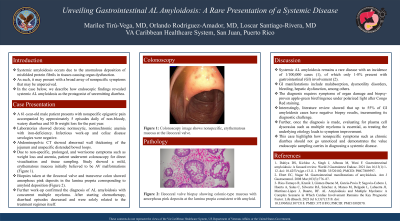
Introduction: Systemic amyloidosis occurs due to the anomalous deposition of misfolded protein fibrils in tissues causing organ dysfunction. As such, it may present with a broad array of nonspecific symptoms that may be unperceived. In the case below, we describe how endoscopic findings revealed systemic AL amyloidosis as the protagonist of unremitting diarrhea.
Case Description/Methods: A 61-year-old male patient presents with nonspecific epigastric pain accompanied by approximately 5 episodes daily of non-bloody, watery diarrhea and 30 lb weight loss for the past year. Laboratories showed chronic normocytic, normochromic anemia with iron-deficiency. Infectious work-up and celiac disease serologies were negative. Abdominopelvic CT showed abnormal wall thickening of the jejunum and unspecific distended bowel loops. Due to non-specific, prolonged, and worrisome symptoms such as weight loss and anemia, patient underwent colonoscopy for direct visualization and tissue sampling. Study showed a mild, erythematous mucosa initially believed to be AV malformations. Biopsies taken at the ileocecal valve and transverse colon showed amorphous pink deposits in the lamina propria corresponding to amyloid deposition. Further work-up confirmed the diagnosis of AL amyloidosis with concurrent multiple myeloma. After starting chemotherapy, diarrheal episodes decreased and were solely related to the treatment regimen itself.
Discussion: Systemic AL amyloidosis remains a rare disease with an incidence of 1/100,000 cases, of which only 1-8% present with gastrointestinal (GI) disease. GI manifestations include malabsorption, dysmotility disorders, bleeding, hepatic dysfunction, among others. The diagnosis requires symptoms of organ damage and biopsy-proven apple-green birefringence under polarized light after Congo Red staining. Interestingly, literature review showed that up to 55% of cases of GI amyloidosis have negative biopsy results, incrementing its diagnostic challenge. Further, once the diagnosis is made, evaluating for plasma cell dyscrasias such as multiple myeloma is essential, as treating the underlying etiology leads to symptom improvement. This case highlights how nonspecific symptoms such as chronic diarrhea should not go unnoticed and demonstrates the value endoscopic sampling carries in diagnosing a systemic disease.

Disclosures:
Marilee Tirú-Vega, MD, Orlando Rodriguez-Amador, MD, Loscar Santiago-Rivera, MD. P0296 - Unveiling Gastrointestinal AL Amyloidosis: A Rare Presentation of a Systemic Disease, ACG 2024 Annual Scientific Meeting Abstracts. Philadelphia, PA: American College of Gastroenterology.
Marilee Tirú-Vega, MD, Orlando Rodriguez-Amador, MD, Loscar Santiago-Rivera, MD
VA Caribbean Healthcare System, San Juan, Puerto Rico
Introduction: Systemic amyloidosis occurs due to the anomalous deposition of misfolded protein fibrils in tissues causing organ dysfunction. As such, it may present with a broad array of nonspecific symptoms that may be unperceived. In the case below, we describe how endoscopic findings revealed systemic AL amyloidosis as the protagonist of unremitting diarrhea.
Case Description/Methods: A 61-year-old male patient presents with nonspecific epigastric pain accompanied by approximately 5 episodes daily of non-bloody, watery diarrhea and 30 lb weight loss for the past year. Laboratories showed chronic normocytic, normochromic anemia with iron-deficiency. Infectious work-up and celiac disease serologies were negative. Abdominopelvic CT showed abnormal wall thickening of the jejunum and unspecific distended bowel loops. Due to non-specific, prolonged, and worrisome symptoms such as weight loss and anemia, patient underwent colonoscopy for direct visualization and tissue sampling. Study showed a mild, erythematous mucosa initially believed to be AV malformations. Biopsies taken at the ileocecal valve and transverse colon showed amorphous pink deposits in the lamina propria corresponding to amyloid deposition. Further work-up confirmed the diagnosis of AL amyloidosis with concurrent multiple myeloma. After starting chemotherapy, diarrheal episodes decreased and were solely related to the treatment regimen itself.
Discussion: Systemic AL amyloidosis remains a rare disease with an incidence of 1/100,000 cases, of which only 1-8% present with gastrointestinal (GI) disease. GI manifestations include malabsorption, dysmotility disorders, bleeding, hepatic dysfunction, among others. The diagnosis requires symptoms of organ damage and biopsy-proven apple-green birefringence under polarized light after Congo Red staining. Interestingly, literature review showed that up to 55% of cases of GI amyloidosis have negative biopsy results, incrementing its diagnostic challenge. Further, once the diagnosis is made, evaluating for plasma cell dyscrasias such as multiple myeloma is essential, as treating the underlying etiology leads to symptom improvement. This case highlights how nonspecific symptoms such as chronic diarrhea should not go unnoticed and demonstrates the value endoscopic sampling carries in diagnosing a systemic disease.

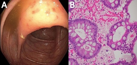
Figure: Figure 1A: Colonoscopy image shows nonspecific, erythematous mucosa at the ileocecal valve.
Figure 1B: Ileocecal valve biopsy showing colonic-type mucosa with amorphous pink deposits at the lamina propia consistent with amyloid.
Figure 1B: Ileocecal valve biopsy showing colonic-type mucosa with amorphous pink deposits at the lamina propia consistent with amyloid.
Disclosures:
Marilee Tirú-Vega indicated no relevant financial relationships.
Orlando Rodriguez-Amador indicated no relevant financial relationships.
Loscar Santiago-Rivera indicated no relevant financial relationships.
Marilee Tirú-Vega, MD, Orlando Rodriguez-Amador, MD, Loscar Santiago-Rivera, MD. P0296 - Unveiling Gastrointestinal AL Amyloidosis: A Rare Presentation of a Systemic Disease, ACG 2024 Annual Scientific Meeting Abstracts. Philadelphia, PA: American College of Gastroenterology.

