Sunday Poster Session
Category: Colon
P0326 - Alone but Not Alone: Isolated Gastrointestinal Amyloidosis
Sunday, October 27, 2024
3:30 PM - 7:00 PM ET
Location: Exhibit Hall E

Has Audio
- KT
Kimberly Tena Diaz, DO
Cooper University Hospital
Camden, NJ
Presenting Author(s)
Kimberly Tena Diaz, DO1, Michael Schwartz, MD2, Adib Chaaya, MD2, Upasana Joneja, MD1
1Cooper University Hospital, Camden, NJ; 2Cooper Health Gastroenterology, Camden, NJ
Introduction: Amyloidosis is characterized by aggregation of insoluble, misfolded proteins that deposit extracellularly leading to tissue dysfunction. There are many amyloid types including an epidermal growth factor-containing fibulin-like extracellular matrix protein 1 (EFEMP1) which causes an age-related venous amyloidosis in the gastrointestinal tract of elder women. This protein type has been identified in patients biopsied for various reasons, such as in our patient. It continues to be rare, with its clinical significance and management guidelines unclear.
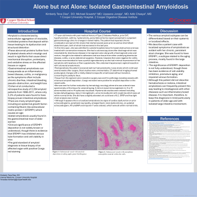
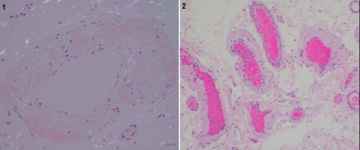
Case Description/Methods: 77-year-old female with past medical history of hysterectomy and periumbilical hernia repairs presented to outside hospitals twice for bowel obstructions and change in stool caliber. She was treated conservatively and her pencil-thin stools were attributed to her obstruction. A colonoscopy done after discharge revealed diverticulosis and a fixed sigmoid colon and rectum, thought to be related to adhesions from either underlying diverticular disease, colitis, or prior abdominal surgeries. She initiated laxatives and fiber supplements for symptom management. However, her symptoms continued, so she presented to outpatient gastroenterology. She was recommended to have a partial sigmoidectomy as she had minimal improvement. She underwent laparoscopic sigmoid resection with colorectal anastomosis. Tissue pathology revealed vessels with intramural amyloid deposition and positive Congo red staining. She was sent for hematology-oncology evaluation which revealed no evidence of plasma cell dyscrasias or signs indicating systemic involvement. Mass spectrometry revealed EFEMP1 amyloidosis.
Discussion: The various amyloid subtypes can be differentiated based on their systemic or localized effects. We describe a patient case with localized symptoms of amyloidosis as evident with her chronic, persistent stool changes. She was found to have EFEMP1, a subtype related to the aging process, mostly found in the large intestine. The significance of EFEMP1 deposition is not fully understood, though there has been evidence of cell viability inhibition, premature aging, and impaired venous formation. Although this patient did not describe hematochezia or melena, intestinal amyloidosis can frequently present this way leading to misdiagnosis with other diseases such as inflammatory bowel disease. It is important, therefore, to keep this diagnosis in mind particularly in patients of older age and with isolated large intestine involvement.

Disclosures:
Kimberly Tena Diaz, DO1, Michael Schwartz, MD2, Adib Chaaya, MD2, Upasana Joneja, MD1. P0326 - Alone but Not Alone: Isolated Gastrointestinal Amyloidosis, ACG 2024 Annual Scientific Meeting Abstracts. Philadelphia, PA: American College of Gastroenterology.
1Cooper University Hospital, Camden, NJ; 2Cooper Health Gastroenterology, Camden, NJ
Introduction: Amyloidosis is characterized by aggregation of insoluble, misfolded proteins that deposit extracellularly leading to tissue dysfunction. There are many amyloid types including an epidermal growth factor-containing fibulin-like extracellular matrix protein 1 (EFEMP1) which causes an age-related venous amyloidosis in the gastrointestinal tract of elder women. This protein type has been identified in patients biopsied for various reasons, such as in our patient. It continues to be rare, with its clinical significance and management guidelines unclear.
Case Description/Methods: 77-year-old female with past medical history of hysterectomy and periumbilical hernia repairs presented to outside hospitals twice for bowel obstructions and change in stool caliber. She was treated conservatively and her pencil-thin stools were attributed to her obstruction. A colonoscopy done after discharge revealed diverticulosis and a fixed sigmoid colon and rectum, thought to be related to adhesions from either underlying diverticular disease, colitis, or prior abdominal surgeries. She initiated laxatives and fiber supplements for symptom management. However, her symptoms continued, so she presented to outpatient gastroenterology. She was recommended to have a partial sigmoidectomy as she had minimal improvement. She underwent laparoscopic sigmoid resection with colorectal anastomosis. Tissue pathology revealed vessels with intramural amyloid deposition and positive Congo red staining. She was sent for hematology-oncology evaluation which revealed no evidence of plasma cell dyscrasias or signs indicating systemic involvement. Mass spectrometry revealed EFEMP1 amyloidosis.
Discussion: The various amyloid subtypes can be differentiated based on their systemic or localized effects. We describe a patient case with localized symptoms of amyloidosis as evident with her chronic, persistent stool changes. She was found to have EFEMP1, a subtype related to the aging process, mostly found in the large intestine. The significance of EFEMP1 deposition is not fully understood, though there has been evidence of cell viability inhibition, premature aging, and impaired venous formation. Although this patient did not describe hematochezia or melena, intestinal amyloidosis can frequently present this way leading to misdiagnosis with other diseases such as inflammatory bowel disease. It is important, therefore, to keep this diagnosis in mind particularly in patients of older age and with isolated large intestine involvement.

Figure: Figure 1: Submucosal vessel with amyloid deposition revealing apple green birefringence
Figure 2: Submucosal vessels with amorphous eosinophilic material within the walls
Figure 2: Submucosal vessels with amorphous eosinophilic material within the walls
Disclosures:
Kimberly Tena Diaz indicated no relevant financial relationships.
Michael Schwartz indicated no relevant financial relationships.
Adib Chaaya indicated no relevant financial relationships.
Upasana Joneja indicated no relevant financial relationships.
Kimberly Tena Diaz, DO1, Michael Schwartz, MD2, Adib Chaaya, MD2, Upasana Joneja, MD1. P0326 - Alone but Not Alone: Isolated Gastrointestinal Amyloidosis, ACG 2024 Annual Scientific Meeting Abstracts. Philadelphia, PA: American College of Gastroenterology.
