Sunday Poster Session
Category: Colon
P0335 - A Case of Encapsulating Peritoneal Sclerosis Complicated by a Large Loculated Fluid Collection
Sunday, October 27, 2024
3:30 PM - 7:00 PM ET
Location: Exhibit Hall E

Has Audio

Rohan Tripathi, BS
Saint Louis University School of Medicine
Phoenix, AZ
Presenting Author(s)
Rohan Tripathi, BS1, Forest Riekhof, BS, MD2, Farzana Hoque, MD, MRCP3
1Saint Louis University School of Medicine, Phoenix, AZ; 2University of Utah School of Medicine, Salt Lake City, UT; 3SSM Health Saint Louis University Hospital, St. Louis, MO
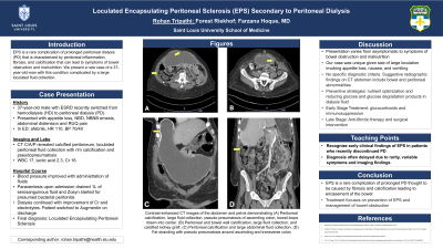
Introduction: Encapsulating peritoneal sclerosis (EPS) is a rare complication of prolonged peritoneal dialysis (PD) that is characterized by peritoneal inflammation, fibrosis, and calcification that can lead to encasement of the bowel leading to symptoms of bowel obstruction and malnutrition. We present a rare case of a 37-year-old-man with this rare condition complicated by a large loculated fluid collection.
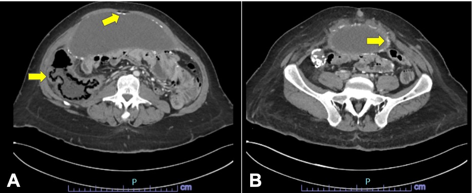
Case Description/Methods: A 37-year-old-man with a history of end-stage-renal disease (ESRD) secondary to congenital obstructive uropathy, who was recently switched to hemodialysis (HD) after 5 years of peritoneal dialysis (PD), presented to the emergency department. They reported 1 to 2 weeks of anorexia, non-bloody diarrhea, non-bloody non-bilious emesis, abdominal distention, and right upper quadrant abdominal pain. Vitals signs revealed tachycardia at 110 beats per minute and hypotension at 70/40 which resolved after administration of fluids. Laboratory parameters significant for leukocytosis of 17, lactic acidosis of 2.3, elevated creatinine of 16, and Ca of 10.0, and phosphorus of 7.2. A CT C/A/P was significant for calcified peritoneum, a large loculated peritoneal fluid collection with rim calcification, mild colonic wall thickening/fat stranding with pseudo pneumatosis and no evidence of acute bowel obstruction (Figure 1). Paracentesis drained 1 liter of serosanguinous fluid upon admission. Dialysis continued with improvement of creatinine and electrolytes.
Discussion: The patient developed a large fluid collection in the setting of encapsulating peritoneal sclerosis, a life-threatening complication of long-term peritoneal dialysis. This case is uncommon due to the size of the large loculation of peritoneal fluid surrounding the thickened peritoneum contributing to encapsulation of the bowel, invoking anorexia, nausea, and vomiting. Diagnosis remains challenging due to diagnostic criteria that rely largely on clinical symptoms and variable radiographic findings. Treatment based on staging has been proposed with emphasis on steroids and immunosuppression in the early stages, and anti-fibrotic therapy and surgical intervention in later stages.

Disclosures:
Rohan Tripathi, BS1, Forest Riekhof, BS, MD2, Farzana Hoque, MD, MRCP3. P0335 - A Case of Encapsulating Peritoneal Sclerosis Complicated by a Large Loculated Fluid Collection, ACG 2024 Annual Scientific Meeting Abstracts. Philadelphia, PA: American College of Gastroenterology.
1Saint Louis University School of Medicine, Phoenix, AZ; 2University of Utah School of Medicine, Salt Lake City, UT; 3SSM Health Saint Louis University Hospital, St. Louis, MO
Introduction: Encapsulating peritoneal sclerosis (EPS) is a rare complication of prolonged peritoneal dialysis (PD) that is characterized by peritoneal inflammation, fibrosis, and calcification that can lead to encasement of the bowel leading to symptoms of bowel obstruction and malnutrition. We present a rare case of a 37-year-old-man with this rare condition complicated by a large loculated fluid collection.
Case Description/Methods: A 37-year-old-man with a history of end-stage-renal disease (ESRD) secondary to congenital obstructive uropathy, who was recently switched to hemodialysis (HD) after 5 years of peritoneal dialysis (PD), presented to the emergency department. They reported 1 to 2 weeks of anorexia, non-bloody diarrhea, non-bloody non-bilious emesis, abdominal distention, and right upper quadrant abdominal pain. Vitals signs revealed tachycardia at 110 beats per minute and hypotension at 70/40 which resolved after administration of fluids. Laboratory parameters significant for leukocytosis of 17, lactic acidosis of 2.3, elevated creatinine of 16, and Ca of 10.0, and phosphorus of 7.2. A CT C/A/P was significant for calcified peritoneum, a large loculated peritoneal fluid collection with rim calcification, mild colonic wall thickening/fat stranding with pseudo pneumatosis and no evidence of acute bowel obstruction (Figure 1). Paracentesis drained 1 liter of serosanguinous fluid upon admission. Dialysis continued with improvement of creatinine and electrolytes.
Discussion: The patient developed a large fluid collection in the setting of encapsulating peritoneal sclerosis, a life-threatening complication of long-term peritoneal dialysis. This case is uncommon due to the size of the large loculation of peritoneal fluid surrounding the thickened peritoneum contributing to encapsulation of the bowel, invoking anorexia, nausea, and vomiting. Diagnosis remains challenging due to diagnostic criteria that rely largely on clinical symptoms and variable radiographic findings. Treatment based on staging has been proposed with emphasis on steroids and immunosuppression in the early stages, and anti-fibrotic therapy and surgical intervention in later stages.

Figure: Figure 1. (A) CT abdomen axial cut showing peritoneal calcification, large fluid collection, pseudo-pneumatosis of ascending colon, bowel loops drawn into center. (B) CT abdomen axial cut showing peritoneal and bowel wall calcification, large fluid collection, and calcified kidney graft.
Disclosures:
Rohan Tripathi indicated no relevant financial relationships.
Forest Riekhof indicated no relevant financial relationships.
Farzana Hoque indicated no relevant financial relationships.
Rohan Tripathi, BS1, Forest Riekhof, BS, MD2, Farzana Hoque, MD, MRCP3. P0335 - A Case of Encapsulating Peritoneal Sclerosis Complicated by a Large Loculated Fluid Collection, ACG 2024 Annual Scientific Meeting Abstracts. Philadelphia, PA: American College of Gastroenterology.
