Sunday Poster Session
Category: Colon
P0342 - Sevelamer Associated Colitis as a Colon Cancer Mimicker
Sunday, October 27, 2024
3:30 PM - 7:00 PM ET
Location: Exhibit Hall E

Has Audio
.jpg)
Brittany Doll, MD
Rush University Medical Center
Chicago, IL
Presenting Author(s)
Brittany Doll, MD, Chandler Shapiro, MD, Zoë Post, MD, Farnaz Shariati, MD, Ram Al-Sabti, MBChB
Rush University Medical Center, Chicago, IL
Introduction: Sevelamer is a non-absorbable resin that binds to phosphate in the gastrointestinal (GI) tract to allow for excretion in stool. It is primarily used to prevent renal osteodystrophy in patients with chronic kidney disease (CKD) and end stage renal disease (ESRD). Prior clinical trials demonstrate that sevelamer is generally well tolerated without adverse GI effects.
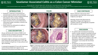
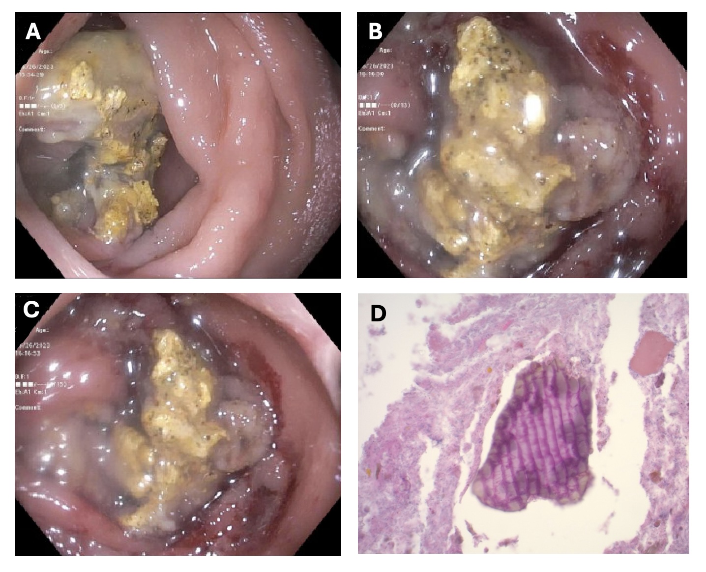
Case Description/Methods: A 40-year-old female with a history of gastric arteriovenous malformation (AVM), ESRD on hemodialysis, opioid-induced constipation, and HIV (CD4 count: 439 cells/uL) presented with acute microcytic on chronic normocytic anemia. On admission, hemoglobin (Hgb) was 10.0 g/dl and mean corpuscular volume (MCV) was 77.3 fL; however, Hgb decreased to 6.8 g/dl over the next 15 days. The patient denied GI symptoms, and there were no overt signs of bleeding. A computed tomography (CT) scan demonstrated short segment mural thickening of the proximal ascending colon and cecum. To further evaluate the etiology of the anemia and CT findings, the patient underwent a colonoscopy which revealed a friable, mass-like lesion with congested, erythematous, and ulcerated mucosa in the cecum. The mass was highly concerning for malignancy based on endoscopic appearance, but pathology showed mild inflammation and regenerative changes with fragments of crystalline material, morphologically compatible with sevelamer crystals. Sevelamer was subsequently discontinued. Outpatient surveillance colonoscopy was scheduled in one month to reassess the site after cessation of sevelamer, but the patient was lost to follow up.
Discussion: A consequence of non-absorbable resins is the ability to crystallize and form concentrations that deposit into the GI mucosa and promote luminal damage. Most commonly, this causes ulcerations and mucosal inflammation. It is rare for sevelamer injury to present as an occult GI bleed with a mass-like colonic lesion that can be mistaken as a malignancy on endoscopic or cross-sectional imaging.
In patients with CKD who develop worsening anemia, it may be reasonable to attribute this to progression of kidney disease especially in the absence of GI symptoms or overt bleeding. However, our case emphasizes that sevelamer-crystal induced mucosal injury can be a source of occult blood loss in this patient population.

Disclosures:
Brittany Doll, MD, Chandler Shapiro, MD, Zoë Post, MD, Farnaz Shariati, MD, Ram Al-Sabti, MBChB. P0342 - Sevelamer Associated Colitis as a Colon Cancer Mimicker, ACG 2024 Annual Scientific Meeting Abstracts. Philadelphia, PA: American College of Gastroenterology.
Rush University Medical Center, Chicago, IL
Introduction: Sevelamer is a non-absorbable resin that binds to phosphate in the gastrointestinal (GI) tract to allow for excretion in stool. It is primarily used to prevent renal osteodystrophy in patients with chronic kidney disease (CKD) and end stage renal disease (ESRD). Prior clinical trials demonstrate that sevelamer is generally well tolerated without adverse GI effects.
Case Description/Methods: A 40-year-old female with a history of gastric arteriovenous malformation (AVM), ESRD on hemodialysis, opioid-induced constipation, and HIV (CD4 count: 439 cells/uL) presented with acute microcytic on chronic normocytic anemia. On admission, hemoglobin (Hgb) was 10.0 g/dl and mean corpuscular volume (MCV) was 77.3 fL; however, Hgb decreased to 6.8 g/dl over the next 15 days. The patient denied GI symptoms, and there were no overt signs of bleeding. A computed tomography (CT) scan demonstrated short segment mural thickening of the proximal ascending colon and cecum. To further evaluate the etiology of the anemia and CT findings, the patient underwent a colonoscopy which revealed a friable, mass-like lesion with congested, erythematous, and ulcerated mucosa in the cecum. The mass was highly concerning for malignancy based on endoscopic appearance, but pathology showed mild inflammation and regenerative changes with fragments of crystalline material, morphologically compatible with sevelamer crystals. Sevelamer was subsequently discontinued. Outpatient surveillance colonoscopy was scheduled in one month to reassess the site after cessation of sevelamer, but the patient was lost to follow up.
Discussion: A consequence of non-absorbable resins is the ability to crystallize and form concentrations that deposit into the GI mucosa and promote luminal damage. Most commonly, this causes ulcerations and mucosal inflammation. It is rare for sevelamer injury to present as an occult GI bleed with a mass-like colonic lesion that can be mistaken as a malignancy on endoscopic or cross-sectional imaging.
In patients with CKD who develop worsening anemia, it may be reasonable to attribute this to progression of kidney disease especially in the absence of GI symptoms or overt bleeding. However, our case emphasizes that sevelamer-crystal induced mucosal injury can be a source of occult blood loss in this patient population.

Figure: Figure 1. Colonoscopy images: (A) Normal appearing mucosa surrounding the cecal mass, (B) and (C) the friable mass-like lesion as seen with surrounding congested, erythematous, and ulcerated mucosa in the cecum. Cecal mass biopsy: (D) histopathology demonstrates the characteristic fish scale pattern associated with sevelamer crystal deposition.
Disclosures:
Brittany Doll indicated no relevant financial relationships.
Chandler Shapiro indicated no relevant financial relationships.
Zoë Post indicated no relevant financial relationships.
Farnaz Shariati indicated no relevant financial relationships.
Ram Al-Sabti indicated no relevant financial relationships.
Brittany Doll, MD, Chandler Shapiro, MD, Zoë Post, MD, Farnaz Shariati, MD, Ram Al-Sabti, MBChB. P0342 - Sevelamer Associated Colitis as a Colon Cancer Mimicker, ACG 2024 Annual Scientific Meeting Abstracts. Philadelphia, PA: American College of Gastroenterology.
