Sunday Poster Session
Category: Colon
P0372 - A Case of Hematochezia Secondary to a Colonic Glomus Tumor
Sunday, October 27, 2024
3:30 PM - 7:00 PM ET
Location: Exhibit Hall E

Has Audio

Jiteshwar S. Pannu, MD
University of Oklahoma Health Sciences Center
Oklahoma City, OK
Presenting Author(s)
Jiteshwar S. Pannu, MD, Luke Sorensen, MD, Andrea Fernandez, MD, Mohammad Madhoun, MD, MS
University of Oklahoma Health Sciences Center, Oklahoma City, OK
Introduction: Glomus tumors (GTs) are rare mesenchymal neoplasms, arising from smooth muscle cells of the thermoregulatory glomus body. While majority of the gastrointestinal (GI) GTs occur in the stomach, colonic GTs are extremely rare. Herein, we present a case of hematochezia, secondary to a colonic GT, that was managed surgically.
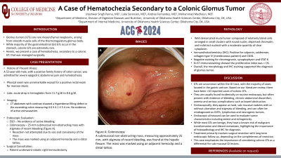
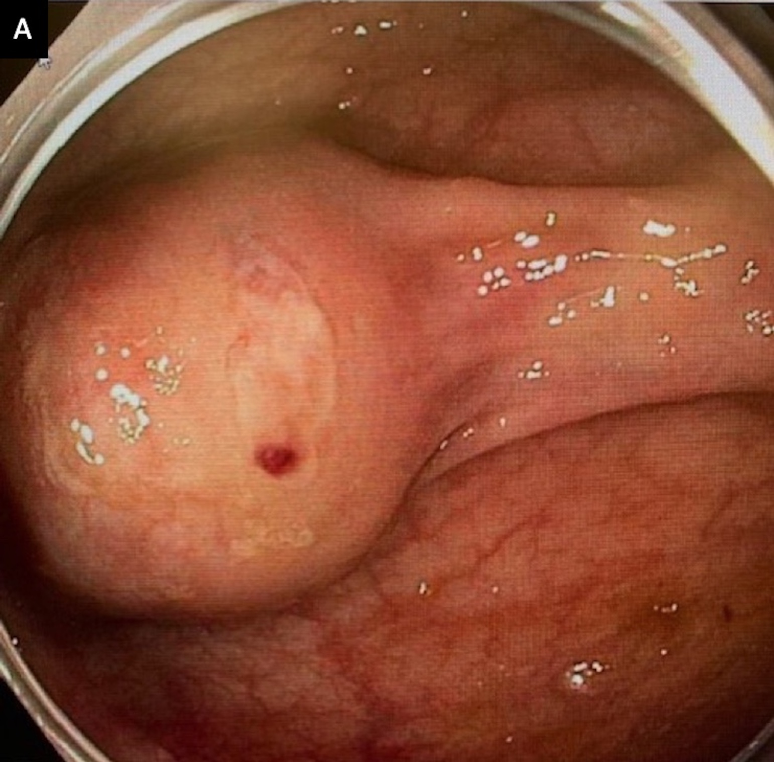
Case Description/Methods: A 52-year-old male with a positive family history of colon cancer, was admitted for severe epigastric abdominal pain and hematochezia. Physical exam was unremarkable except a positive rectal exam for maroon stools. Bloodwork revealed an acute drop in hemoglobin from 11.7 g/dl to 8.6 g/dl. CT abdomen with contrast showed hyperdense filling defect in the ascending colon measuring 4 X 4.1 X 5.4 cm. He was evaluated with an EGD that showed no identifiable source of bleeding. Therefore, a colonoscopy was performed, revealing a sub-mucosal, non-obstructing mass at the hepatic flexure with stigmata of recent bleeding, suspicious for GIST. The mass was marked using an adjacent hemoclip and a distal tattoo. He subsequently underwent a robotic right hemicolectomy. Histo-pathological (HP) and gross examination revealed a well demarcated mural tumor with relatively bland cells in clusters, round nuclei, well dispersed chromatin, indistinct nucleoli and clear cytoplasm. Immunohistochemistry (IHC) was positive for calponin, caldesmon, collagen IV and CD34 supporting the diagnosis of a primary colonic GT.
Discussion: GTs are uncommon within the GI tract, with the majority of cases located in the gastric antrum. Based on our literature review, there have been < 10 reported cases of colonic GTs. They are usually found incidentally on routine endoscopy, but often present with evidence of bleeding, chronic abdominal discomfort, anemia and serious complications such as bowel obstruction. Endoscopically, they appear as hard, sub-mucosal nodules with or without ulceration and stigmata of bleeding, and can often be misdiagnosed as GISTs, lymphomas and neurogenic tumors. Endoscopic ultrasound can be used to evaluate tumor characteristics including extent and echogenicity. While most GTs are benign, they have a known risk of malignant transformation and distant metastasis, highlighting the importance of HP and IHC examination for diagnosis. Treatment primarily involves surgical resection with long term endoscopic follow up, depending on the completeness of resection. This case emphasizes the importance of considering colonic GTs as a differential for sub-mucosal GI lesions.

Disclosures:
Jiteshwar S. Pannu, MD, Luke Sorensen, MD, Andrea Fernandez, MD, Mohammad Madhoun, MD, MS. P0372 - A Case of Hematochezia Secondary to a Colonic Glomus Tumor, ACG 2024 Annual Scientific Meeting Abstracts. Philadelphia, PA: American College of Gastroenterology.
University of Oklahoma Health Sciences Center, Oklahoma City, OK
Introduction: Glomus tumors (GTs) are rare mesenchymal neoplasms, arising from smooth muscle cells of the thermoregulatory glomus body. While majority of the gastrointestinal (GI) GTs occur in the stomach, colonic GTs are extremely rare. Herein, we present a case of hematochezia, secondary to a colonic GT, that was managed surgically.
Case Description/Methods: A 52-year-old male with a positive family history of colon cancer, was admitted for severe epigastric abdominal pain and hematochezia. Physical exam was unremarkable except a positive rectal exam for maroon stools. Bloodwork revealed an acute drop in hemoglobin from 11.7 g/dl to 8.6 g/dl. CT abdomen with contrast showed hyperdense filling defect in the ascending colon measuring 4 X 4.1 X 5.4 cm. He was evaluated with an EGD that showed no identifiable source of bleeding. Therefore, a colonoscopy was performed, revealing a sub-mucosal, non-obstructing mass at the hepatic flexure with stigmata of recent bleeding, suspicious for GIST. The mass was marked using an adjacent hemoclip and a distal tattoo. He subsequently underwent a robotic right hemicolectomy. Histo-pathological (HP) and gross examination revealed a well demarcated mural tumor with relatively bland cells in clusters, round nuclei, well dispersed chromatin, indistinct nucleoli and clear cytoplasm. Immunohistochemistry (IHC) was positive for calponin, caldesmon, collagen IV and CD34 supporting the diagnosis of a primary colonic GT.
Discussion: GTs are uncommon within the GI tract, with the majority of cases located in the gastric antrum. Based on our literature review, there have been < 10 reported cases of colonic GTs. They are usually found incidentally on routine endoscopy, but often present with evidence of bleeding, chronic abdominal discomfort, anemia and serious complications such as bowel obstruction. Endoscopically, they appear as hard, sub-mucosal nodules with or without ulceration and stigmata of bleeding, and can often be misdiagnosed as GISTs, lymphomas and neurogenic tumors. Endoscopic ultrasound can be used to evaluate tumor characteristics including extent and echogenicity. While most GTs are benign, they have a known risk of malignant transformation and distant metastasis, highlighting the importance of HP and IHC examination for diagnosis. Treatment primarily involves surgical resection with long term endoscopic follow up, depending on the completeness of resection. This case emphasizes the importance of considering colonic GTs as a differential for sub-mucosal GI lesions.

Figure: Figure A: sub-mucosal, non-obstructing mass at the hepatic flexure with stigmata of recent bleeding
Disclosures:
Jiteshwar Pannu indicated no relevant financial relationships.
Luke Sorensen indicated no relevant financial relationships.
Andrea Fernandez indicated no relevant financial relationships.
Mohammad Madhoun indicated no relevant financial relationships.
Jiteshwar S. Pannu, MD, Luke Sorensen, MD, Andrea Fernandez, MD, Mohammad Madhoun, MD, MS. P0372 - A Case of Hematochezia Secondary to a Colonic Glomus Tumor, ACG 2024 Annual Scientific Meeting Abstracts. Philadelphia, PA: American College of Gastroenterology.
