Sunday Poster Session
Category: Colon
P0374 - Large Tubulovillous Rectal Mass Causing McKittrick-Wheelock Syndrome (MWS)
Sunday, October 27, 2024
3:30 PM - 7:00 PM ET
Location: Exhibit Hall E

Has Audio

Danzhu Zhao, DO
University of Connecticut Health Center
WEST HARTFORD, CT
Presenting Author(s)
Danzhu Zhao, DO1, Bianca Thakkar, DO1, Teresa Da Cunha, MD1, Minh Thu T.. Nguyen, MD1, Alexandre Vdovenko, MD2, Alexander Potashinsky, MD3
1University of Connecticut Health Center, Farmington, CT; 2Hartford Pathology Associates, PC, Hospital of Central Connecticut, New Britain, CT; 3Hartford Healthcare, New Britain, CT
Introduction: Villous adenomas can cause severe electrolyte imbalances and acute kidney injury due to secretory diarrhea, known as McKittrick-Wheelock Syndrome (MWS). While MWS is documented in villous adenomas, literature on MWS from tubulovillous adenomas, as in our case, is limited.
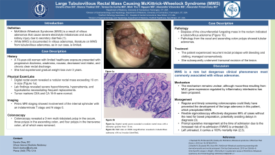
Case Description/Methods: A 74-year-old woman with limited healthcare exposure presented with progressive dizziness, weakness, nausea, decreased oral intake, and chronic clear rectal discharge. She had experienced gradual weight loss over 3 years. Lab findings revealed severe hypochloremia, hyponatremia, and hypokalemia necessitating frequent replacements. Tumor markers CEA and CA 19-9 were normal. Abdominal and pelvic CT imaging showed a distended rectum measuring 9.2 cm by 8.8 cm with mixed fluid and dense soft tissue-like material merging with the rectal wall, raising concern for a rectal neoplasm. Digital rectal exam revealed a nodular rectal mass exceeding 10 cm in size (Figure 1a).
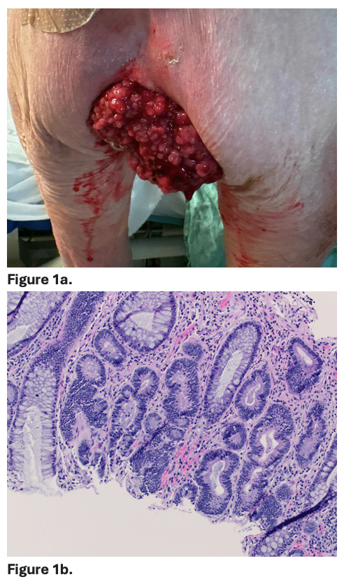
A few days after admission, colonoscopy revealed a 9 cm multi-lobulated polyp in the cecum, three polyps in the ascending colon, and four polyps in the transverse colon, all of which were removed. Biopsies of the circumferential fungating mass in the rectum indicated a tubulovillous adenoma (Figure 1b). Pathology from the cecal and ascending colon polyps showed tubular adenomas. Pelvic MRI staging showed involvement of the internal sphincter with an indeterminate T stage and N stage 0. During her hospital stay, the patient experienced recurrent rectal prolapse with bleeding and clotting, managed conservatively. She subsequently underwent transanal excision of the lesion. Final pathology results are pending.
Discussion: MWS is a rare but dangerous clinical phenomenon most commonly associated with villous adenomas. Left untreated, it carries a significant mortality risk. The mechanism remains unclear, although mucorrhea resulting from MUC gene expression regulated by inflammatory interleukins has been proposed. Regular and timely screening colonoscopies could likely have prevented the development of the large adenoma in this patient, which precipitated MWS. Orchard et al. suggested that flexible sigmoidoscopy can effectively diagnose 99% of cases without the need for bowel preparation, potentially avoiding delays in diagnosis associated with colonoscopies. They also recommended prompt operative management at the time of admission due to the increased risk of re-admission if MWS is untreated.

Disclosures:
Danzhu Zhao, DO1, Bianca Thakkar, DO1, Teresa Da Cunha, MD1, Minh Thu T.. Nguyen, MD1, Alexandre Vdovenko, MD2, Alexander Potashinsky, MD3. P0374 - Large Tubulovillous Rectal Mass Causing McKittrick-Wheelock Syndrome (MWS), ACG 2024 Annual Scientific Meeting Abstracts. Philadelphia, PA: American College of Gastroenterology.
1University of Connecticut Health Center, Farmington, CT; 2Hartford Pathology Associates, PC, Hospital of Central Connecticut, New Britain, CT; 3Hartford Healthcare, New Britain, CT
Introduction: Villous adenomas can cause severe electrolyte imbalances and acute kidney injury due to secretory diarrhea, known as McKittrick-Wheelock Syndrome (MWS). While MWS is documented in villous adenomas, literature on MWS from tubulovillous adenomas, as in our case, is limited.
Case Description/Methods: A 74-year-old woman with limited healthcare exposure presented with progressive dizziness, weakness, nausea, decreased oral intake, and chronic clear rectal discharge. She had experienced gradual weight loss over 3 years. Lab findings revealed severe hypochloremia, hyponatremia, and hypokalemia necessitating frequent replacements. Tumor markers CEA and CA 19-9 were normal. Abdominal and pelvic CT imaging showed a distended rectum measuring 9.2 cm by 8.8 cm with mixed fluid and dense soft tissue-like material merging with the rectal wall, raising concern for a rectal neoplasm. Digital rectal exam revealed a nodular rectal mass exceeding 10 cm in size (Figure 1a).
A few days after admission, colonoscopy revealed a 9 cm multi-lobulated polyp in the cecum, three polyps in the ascending colon, and four polyps in the transverse colon, all of which were removed. Biopsies of the circumferential fungating mass in the rectum indicated a tubulovillous adenoma (Figure 1b). Pathology from the cecal and ascending colon polyps showed tubular adenomas. Pelvic MRI staging showed involvement of the internal sphincter with an indeterminate T stage and N stage 0. During her hospital stay, the patient experienced recurrent rectal prolapse with bleeding and clotting, managed conservatively. She subsequently underwent transanal excision of the lesion. Final pathology results are pending.
Discussion: MWS is a rare but dangerous clinical phenomenon most commonly associated with villous adenomas. Left untreated, it carries a significant mortality risk. The mechanism remains unclear, although mucorrhea resulting from MUC gene expression regulated by inflammatory interleukins has been proposed. Regular and timely screening colonoscopies could likely have prevented the development of the large adenoma in this patient, which precipitated MWS. Orchard et al. suggested that flexible sigmoidoscopy can effectively diagnose 99% of cases without the need for bowel preparation, potentially avoiding delays in diagnosis associated with colonoscopies. They also recommended prompt operative management at the time of admission due to the increased risk of re-admission if MWS is untreated.

Figure: Figure 1a. Digital rectal exam revealed a nodular rectal mass with a diameter greater than 10 cm.
Figure 1b. H&E stain at 200X magnification revealed a tubulovillous adenoma with no invasion identified.
Figure 1b. H&E stain at 200X magnification revealed a tubulovillous adenoma with no invasion identified.
Disclosures:
Danzhu Zhao indicated no relevant financial relationships.
Bianca Thakkar indicated no relevant financial relationships.
Teresa Da Cunha indicated no relevant financial relationships.
Minh Thu Nguyen indicated no relevant financial relationships.
Alexandre Vdovenko indicated no relevant financial relationships.
Alexander Potashinsky indicated no relevant financial relationships.
Danzhu Zhao, DO1, Bianca Thakkar, DO1, Teresa Da Cunha, MD1, Minh Thu T.. Nguyen, MD1, Alexandre Vdovenko, MD2, Alexander Potashinsky, MD3. P0374 - Large Tubulovillous Rectal Mass Causing McKittrick-Wheelock Syndrome (MWS), ACG 2024 Annual Scientific Meeting Abstracts. Philadelphia, PA: American College of Gastroenterology.

