Sunday Poster Session
Category: Colon
P0375 - A Bone to Pick: Dystrophic Ossification in a Colon Polyp
Sunday, October 27, 2024
3:30 PM - 7:00 PM ET
Location: Exhibit Hall E

Has Audio

Danzhu Zhao, DO
University of Connecticut Health Center
WEST HARTFORD, CT
Presenting Author(s)
Danzhu Zhao, DO1, Minh Thu T.. Nguyen, MD1, Bianca Thakkar, DO1, Barry Jacobs, MD2, Alexander Potashinsky, MD3
1University of Connecticut Health Center, Farmington, CT; 2Hartford HealthCare, New Britain, CT; 3Hartford Healthcare, New Britain, CT
Introduction: Dystrophic ossification, also known as heterotopic ossification, refers to the formation of bone in non-skeletal tissues. Although rare, its pathogenesis remains unclear. In certain cases documented in the literature, there is a potential link to malignancy, especially with metaplastic ossification incidentally found in colon polyps.
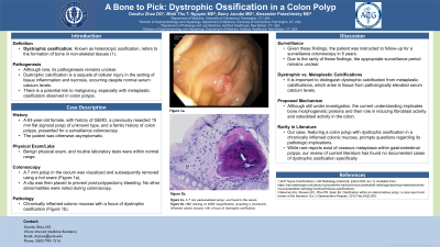
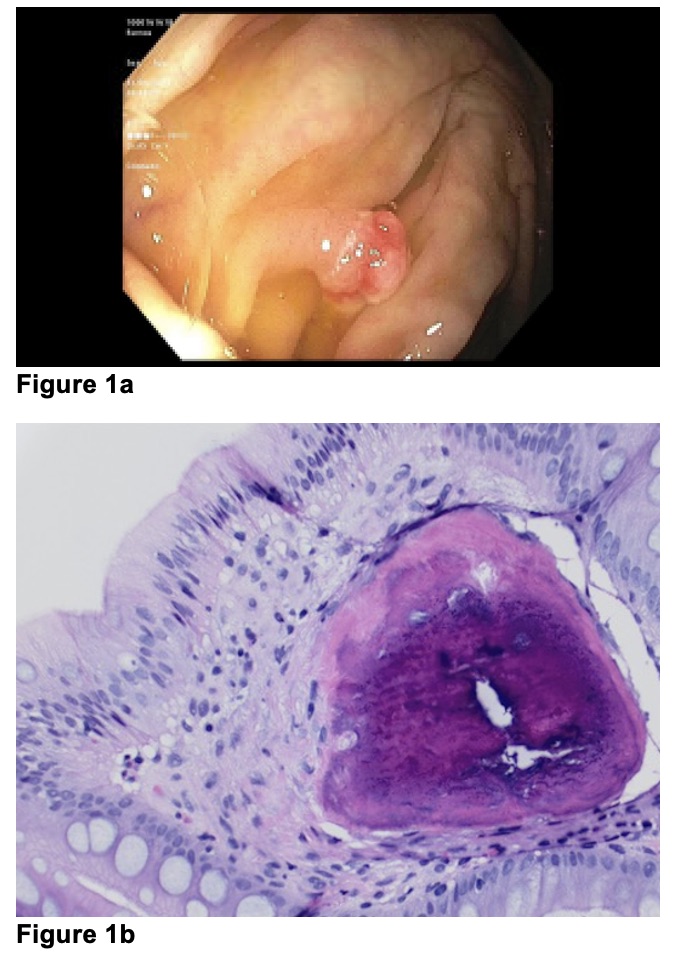
Case Description/Methods: A 63-year-old female with a medical history notable for GERD, a previously resected 15 mm flat sigmoid polyp of unknown type, and a family history of colon polyps, presented for a surveillance colonoscopy. The patient was otherwise asymptomatic with a benign physical exam, and routine laboratory tests were within normal range. During colonoscopy, a 7 mm polyp in the cecum was visualized and subsequently removed using a hot snare (Figure 1a). A clip was then placed to prevent post-polypectomy bleeding. No other abnormalities were noted during colonoscopy. The resected polyp was sent to pathology, revealing a chronically inflamed colonic mucosa with a focus of dystrophic ossification (Figure 1b). Given these findings, the patient was instructed to follow-up for a surveillance colonoscopy in 5 years.
Discussion: Dystrophic calcification is a sequela of cellular injury in the setting of tissue inflammation and necrosis, occurring despite normal serum calcium levels. It is important to distinguish this from metaplastic calcifications, which arise in tissue from pathologically elevated serum calcium levels. While rare reports exist of osseous metaplasia within gastrointestinal polyps, our review of current literature has found no documented cases of dystrophic ossification specifically. Although still under investigation, the current understanding implicates bone morphogenetic proteins and their role in inducing fibroblast activity and osteoblast activity in the colon.
Our case, featuring a colon polyp with dystrophic ossification in a chronically inflamed colonic mucosa, prompts questions regarding its pathologic implications. A 2012 European review identified 22 cases of ossification in colon polyps, revealing that each instance involved advanced polyps. Moreover, approximately half of colonic neoplasms with ossification were associated with colonic malignancy (1). Due to the rarity of these findings, the appropriate surveillance period remains unclear.
References:
1. Bowman EA, Stevens EC, Pfau PR, Spier BJ. Ossification within an adenomatous polyp: a case report and review of the literature. Eur J Gastroenterol Hepatol. 2012 Feb;24(2):209.

Disclosures:
Danzhu Zhao, DO1, Minh Thu T.. Nguyen, MD1, Bianca Thakkar, DO1, Barry Jacobs, MD2, Alexander Potashinsky, MD3. P0375 - A Bone to Pick: Dystrophic Ossification in a Colon Polyp, ACG 2024 Annual Scientific Meeting Abstracts. Philadelphia, PA: American College of Gastroenterology.
1University of Connecticut Health Center, Farmington, CT; 2Hartford HealthCare, New Britain, CT; 3Hartford Healthcare, New Britain, CT
Introduction: Dystrophic ossification, also known as heterotopic ossification, refers to the formation of bone in non-skeletal tissues. Although rare, its pathogenesis remains unclear. In certain cases documented in the literature, there is a potential link to malignancy, especially with metaplastic ossification incidentally found in colon polyps.
Case Description/Methods: A 63-year-old female with a medical history notable for GERD, a previously resected 15 mm flat sigmoid polyp of unknown type, and a family history of colon polyps, presented for a surveillance colonoscopy. The patient was otherwise asymptomatic with a benign physical exam, and routine laboratory tests were within normal range. During colonoscopy, a 7 mm polyp in the cecum was visualized and subsequently removed using a hot snare (Figure 1a). A clip was then placed to prevent post-polypectomy bleeding. No other abnormalities were noted during colonoscopy. The resected polyp was sent to pathology, revealing a chronically inflamed colonic mucosa with a focus of dystrophic ossification (Figure 1b). Given these findings, the patient was instructed to follow-up for a surveillance colonoscopy in 5 years.
Discussion: Dystrophic calcification is a sequela of cellular injury in the setting of tissue inflammation and necrosis, occurring despite normal serum calcium levels. It is important to distinguish this from metaplastic calcifications, which arise in tissue from pathologically elevated serum calcium levels. While rare reports exist of osseous metaplasia within gastrointestinal polyps, our review of current literature has found no documented cases of dystrophic ossification specifically. Although still under investigation, the current understanding implicates bone morphogenetic proteins and their role in inducing fibroblast activity and osteoblast activity in the colon.
Our case, featuring a colon polyp with dystrophic ossification in a chronically inflamed colonic mucosa, prompts questions regarding its pathologic implications. A 2012 European review identified 22 cases of ossification in colon polyps, revealing that each instance involved advanced polyps. Moreover, approximately half of colonic neoplasms with ossification were associated with colonic malignancy (1). Due to the rarity of these findings, the appropriate surveillance period remains unclear.
References:
1. Bowman EA, Stevens EC, Pfau PR, Spier BJ. Ossification within an adenomatous polyp: a case report and review of the literature. Eur J Gastroenterol Hepatol. 2012 Feb;24(2):209.

Figure: Figure 1a. A 7 mm pedunculated polyp was found in the cecum.
Figure 1b. H&E staining at 400X magnification revealing a chronically inflamed colonic mucosa with a focus of dystrophic ossification.
Figure 1b. H&E staining at 400X magnification revealing a chronically inflamed colonic mucosa with a focus of dystrophic ossification.
Disclosures:
Danzhu Zhao indicated no relevant financial relationships.
Minh Thu Nguyen indicated no relevant financial relationships.
Bianca Thakkar indicated no relevant financial relationships.
Barry Jacobs indicated no relevant financial relationships.
Alexander Potashinsky indicated no relevant financial relationships.
Danzhu Zhao, DO1, Minh Thu T.. Nguyen, MD1, Bianca Thakkar, DO1, Barry Jacobs, MD2, Alexander Potashinsky, MD3. P0375 - A Bone to Pick: Dystrophic Ossification in a Colon Polyp, ACG 2024 Annual Scientific Meeting Abstracts. Philadelphia, PA: American College of Gastroenterology.
