Sunday Poster Session
Category: Esophagus
P0564 - Razor Blade Ingestion: A Piece-by-Piece Removal Process
Sunday, October 27, 2024
3:30 PM - 7:00 PM ET
Location: Exhibit Hall E

Has Audio

Allison Derise, MD
Louisiana State University Health
Kenner, LA
Presenting Author(s)
Allison Derise, MD1, Jason Stibbe, MD2, Catherine T.. Hudson, MD, MPH3
1Louisiana State University Health, Kenner, LA; 2LSU Health New Orleans School of Medicine, New Orleans, LA; 3LSU, New Orleans, LA
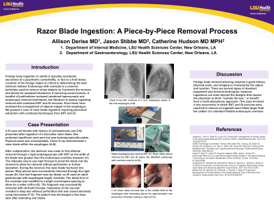
Introduction: Foreign body ingestion in adults is typically accidental, secondary to a psychiatric comorbidity, or due to a food bolus. Location of the foreign object is critical in determining the best retrieval method. Endoscopy with overtube is a common technique used to remove sharp objects as it protects the mucosa and allows for repeated intubations if removing several pieces. A handful of publications reviewed combined laparoscopic and endoscopic removal techniques, but literature is sparse regarding removal with combined ENT and GI services. Even fewer have reviewed the management of objects lodged in the esophagus. We present a case of razor blade ingestion requiring piecemeal extraction with combined techniques from ENT and GI.
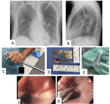
Case Description/Methods: A 61-year-old female with history of schizophrenia and CVA presented after ingestion of a boxcutter razor blade. She endorsed significant neck pain but was hemodynamically stable. Physical exam was unremarkable. Chest X-ray demonstrated a razor blade within the esophagus (A-B).
After collaboration, the decision was made to first attempt removal through a rigid esophagoscope with ENT as the width of the blade was greater than the endoscope overtube diameter (C). The rationale was to use rigid forceps to bend the blade into the channel to allow for removal without perforation or further laceration. During the removal, the razor blade fractured into pieces. Most pieces were successfully removed through the rigid scope (D). One last fragment was too distal, so GI used an adult gastroscope with esophageal length overtube. The distal 2cm of the overtube was modified to a slight oval-shaped opening to allow for a wider orifice (E). The fragment was successfully removed with rat-tooth forceps. Inspection of the mucosa revealed a deep tear without perforation that was closed electively using hemoclips (F-G). The patient was discharged a few days later after tolerating oral intake.
Discussion: Foreign body removal planning requires a good history, physical exam, and imaging to characterize the object and location. There are several types of standard equipment and removal techniques; however, ingestions can arise beyond the designs that require the physician to think “outside the box,” or benefit from a multi-disciplinary approach. This case involved a rare occurrence in which ENT and GI services were required to remove an ingested razor blade larger than the caliber of a standard flexible endoscopic overtube.

Disclosures:
Allison Derise, MD1, Jason Stibbe, MD2, Catherine T.. Hudson, MD, MPH3. P0564 - Razor Blade Ingestion: A Piece-by-Piece Removal Process, ACG 2024 Annual Scientific Meeting Abstracts. Philadelphia, PA: American College of Gastroenterology.
1Louisiana State University Health, Kenner, LA; 2LSU Health New Orleans School of Medicine, New Orleans, LA; 3LSU, New Orleans, LA
Introduction: Foreign body ingestion in adults is typically accidental, secondary to a psychiatric comorbidity, or due to a food bolus. Location of the foreign object is critical in determining the best retrieval method. Endoscopy with overtube is a common technique used to remove sharp objects as it protects the mucosa and allows for repeated intubations if removing several pieces. A handful of publications reviewed combined laparoscopic and endoscopic removal techniques, but literature is sparse regarding removal with combined ENT and GI services. Even fewer have reviewed the management of objects lodged in the esophagus. We present a case of razor blade ingestion requiring piecemeal extraction with combined techniques from ENT and GI.
Case Description/Methods: A 61-year-old female with history of schizophrenia and CVA presented after ingestion of a boxcutter razor blade. She endorsed significant neck pain but was hemodynamically stable. Physical exam was unremarkable. Chest X-ray demonstrated a razor blade within the esophagus (A-B).
After collaboration, the decision was made to first attempt removal through a rigid esophagoscope with ENT as the width of the blade was greater than the endoscope overtube diameter (C). The rationale was to use rigid forceps to bend the blade into the channel to allow for removal without perforation or further laceration. During the removal, the razor blade fractured into pieces. Most pieces were successfully removed through the rigid scope (D). One last fragment was too distal, so GI used an adult gastroscope with esophageal length overtube. The distal 2cm of the overtube was modified to a slight oval-shaped opening to allow for a wider orifice (E). The fragment was successfully removed with rat-tooth forceps. Inspection of the mucosa revealed a deep tear without perforation that was closed electively using hemoclips (F-G). The patient was discharged a few days later after tolerating oral intake.
Discussion: Foreign body removal planning requires a good history, physical exam, and imaging to characterize the object and location. There are several types of standard equipment and removal techniques; however, ingestions can arise beyond the designs that require the physician to think “outside the box,” or benefit from a multi-disciplinary approach. This case involved a rare occurrence in which ENT and GI services were required to remove an ingested razor blade larger than the caliber of a standard flexible endoscopic overtube.

Figure: Figure 1. Chest X-ray with evidence of a 3cm radiopaque object in the mid esophagus (A-B). Rigid esophagoscope used by ENT (C). Razor blade pieces removed by ENT and GI teams (D). Modified endoscope with overtube used by GI (E). 3cm linear deep mucosal tear in the middle third of the esophagus with hemoclips placed for approximation and prevention of further oozing or injury (F-G).
Disclosures:
Allison Derise indicated no relevant financial relationships.
Jason Stibbe indicated no relevant financial relationships.
Catherine Hudson indicated no relevant financial relationships.
Allison Derise, MD1, Jason Stibbe, MD2, Catherine T.. Hudson, MD, MPH3. P0564 - Razor Blade Ingestion: A Piece-by-Piece Removal Process, ACG 2024 Annual Scientific Meeting Abstracts. Philadelphia, PA: American College of Gastroenterology.

