Sunday Poster Session
Category: Esophagus
P0568 - A Rare Case of Esophageal Malignancy Derived From Colonic Origin
Sunday, October 27, 2024
3:30 PM - 7:00 PM ET
Location: Exhibit Hall E

Has Audio

Aarthi Selvam, MD
NYC Health + Hospitals/South Brooklyn Health
Short Hills , NJ
Presenting Author(s)
Aarthi Selvam, MD1, Nikisha Pandya, MD2, Emmanuel Inam, BS2, Joshua Elefteratos, MD1, Michael Bernstein, MD2
1NYC Health + Hospitals/South Brooklyn Health, Brooklyn, NY; 2South Brooklyn Health, Brooklyn, NY
Introduction: There have only been 7 reported cases published in PubMed literature of metastases resulting in esophageal adenocarcinoma from colorectal cancer from 1976 to date. This suggests that esophageal adenocarcinoma from colon metastasis is a rare subset of infrequent entities. Here, we present a unique case of esophageal adenocarcinoma with positive cytokines specific to a tumor source from the epithelium of the lower GI tract in the absence of a cancer diagnosis.
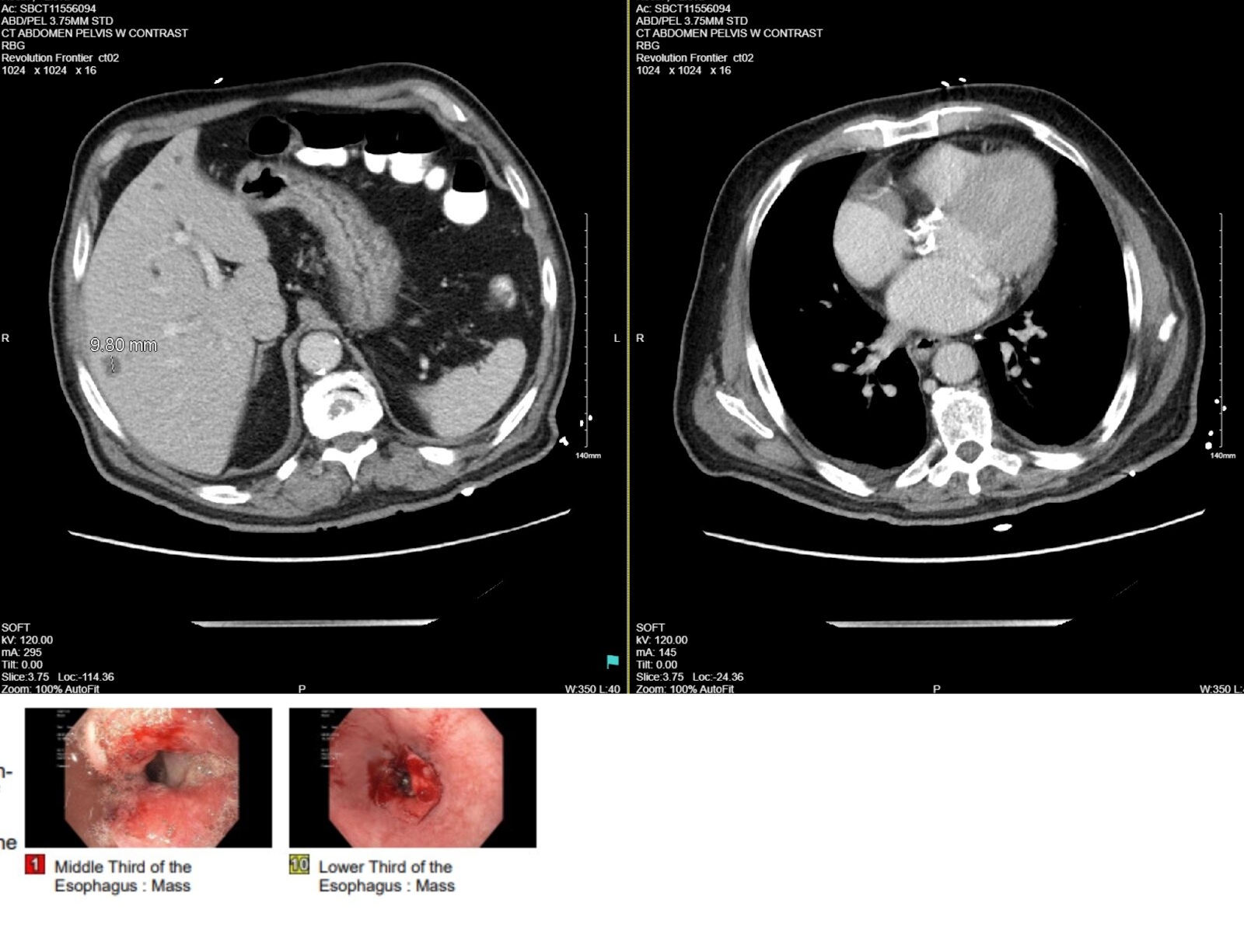
Case Description/Methods: This case presents an 84 y/o M with a history of hypertension, benign prostatic hyperplasia, prostatic cancer, squamous cell carcinoma of the skin, aortic valve stenosis, gastroesophageal reflux disease, and hemorrhoids who was referred to the hospital by his physician after laboratory results confirmed an abnormally low hemoglobin. He had endorsed weakness, fatigue, palpitations and recent unintentional weight loss. Initial laboratory results showed normocytic anemia (Hb: 5.8 g/dl, MCV: 84.2) and elevated reticulocyte count (2%, reference rage: 0.5-1.5%). CT abdomen pelvis with contrast revealed an irregular mass like wall thickening in the region of the distal esophagus near the gastroesophageal junction related to a distal esophageal mass (A). Hepatomegaly with fatty infiltration and multiple cystic lesions within the liver were noted. During EGD, a large, partially obstructing, ulcerating mass involving 1⁄3 of the lumen circumference with friable mucosa was present in the distal esophagus (B).
Surgical pathology was positive for CDX2 and CK20 and negative CK7 reaction. The biopsy of the ulcerated esophageal mass was suggestive of locally advanced non metastatic invasive adenocarcinoma of colonic origin. Colonoscopy showed three benign polyps that were removed.
Discussion: This patient’s esophageal cancer with CK7-/CK20+ immunophenotype points to a colonic origin. However, there was no diagnosis of malignant colorectal carcinoma in this patient, nor was there a history of a prior malignancy. This is a distinction from the aforementioned reported literature cases which had their primary sites of metastases from a malignancy in the lower GI tract. There findings from the pathology report and EGD indicate that this patient’s esophageal tumor was one of primary origin presenting with cytokines of lower GI tract, and not a secondary tumor of distant metastatic spread, making it one of the rarest cases of esophageal carcinoma. Unfortunately, our patient was lost to follow up despite multiple attempts to contact him.

Disclosures:
Aarthi Selvam, MD1, Nikisha Pandya, MD2, Emmanuel Inam, BS2, Joshua Elefteratos, MD1, Michael Bernstein, MD2. P0568 - A Rare Case of Esophageal Malignancy Derived From Colonic Origin, ACG 2024 Annual Scientific Meeting Abstracts. Philadelphia, PA: American College of Gastroenterology.
1NYC Health + Hospitals/South Brooklyn Health, Brooklyn, NY; 2South Brooklyn Health, Brooklyn, NY
Introduction: There have only been 7 reported cases published in PubMed literature of metastases resulting in esophageal adenocarcinoma from colorectal cancer from 1976 to date. This suggests that esophageal adenocarcinoma from colon metastasis is a rare subset of infrequent entities. Here, we present a unique case of esophageal adenocarcinoma with positive cytokines specific to a tumor source from the epithelium of the lower GI tract in the absence of a cancer diagnosis.
Case Description/Methods: This case presents an 84 y/o M with a history of hypertension, benign prostatic hyperplasia, prostatic cancer, squamous cell carcinoma of the skin, aortic valve stenosis, gastroesophageal reflux disease, and hemorrhoids who was referred to the hospital by his physician after laboratory results confirmed an abnormally low hemoglobin. He had endorsed weakness, fatigue, palpitations and recent unintentional weight loss. Initial laboratory results showed normocytic anemia (Hb: 5.8 g/dl, MCV: 84.2) and elevated reticulocyte count (2%, reference rage: 0.5-1.5%). CT abdomen pelvis with contrast revealed an irregular mass like wall thickening in the region of the distal esophagus near the gastroesophageal junction related to a distal esophageal mass (A). Hepatomegaly with fatty infiltration and multiple cystic lesions within the liver were noted. During EGD, a large, partially obstructing, ulcerating mass involving 1⁄3 of the lumen circumference with friable mucosa was present in the distal esophagus (B).
Surgical pathology was positive for CDX2 and CK20 and negative CK7 reaction. The biopsy of the ulcerated esophageal mass was suggestive of locally advanced non metastatic invasive adenocarcinoma of colonic origin. Colonoscopy showed three benign polyps that were removed.
Discussion: This patient’s esophageal cancer with CK7-/CK20+ immunophenotype points to a colonic origin. However, there was no diagnosis of malignant colorectal carcinoma in this patient, nor was there a history of a prior malignancy. This is a distinction from the aforementioned reported literature cases which had their primary sites of metastases from a malignancy in the lower GI tract. There findings from the pathology report and EGD indicate that this patient’s esophageal tumor was one of primary origin presenting with cytokines of lower GI tract, and not a secondary tumor of distant metastatic spread, making it one of the rarest cases of esophageal carcinoma. Unfortunately, our patient was lost to follow up despite multiple attempts to contact him.

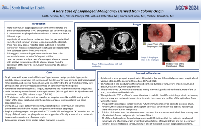
Figure: Image A. Esophageal mass with wall thickening
Image B. Esophageal mass in middle and lower third of the esophagus
Image B. Esophageal mass in middle and lower third of the esophagus
Disclosures:
Aarthi Selvam indicated no relevant financial relationships.
Nikisha Pandya indicated no relevant financial relationships.
Emmanuel Inam indicated no relevant financial relationships.
Joshua Elefteratos indicated no relevant financial relationships.
Michael Bernstein indicated no relevant financial relationships.
Aarthi Selvam, MD1, Nikisha Pandya, MD2, Emmanuel Inam, BS2, Joshua Elefteratos, MD1, Michael Bernstein, MD2. P0568 - A Rare Case of Esophageal Malignancy Derived From Colonic Origin, ACG 2024 Annual Scientific Meeting Abstracts. Philadelphia, PA: American College of Gastroenterology.
