Sunday Poster Session
Category: Esophagus
P0571 - Myopericytoma of the Esophagus: A New Location
Sunday, October 27, 2024
3:30 PM - 7:00 PM ET
Location: Exhibit Hall E

Has Audio

William Abel, MD
Carilion Clinic
Roanoke, VA
Presenting Author(s)
William Abel, MD1, John Rozenberg, MD1, Joel Joseph, MD1, Adil Mir, MD1, Seemeen Hassan, MD1, Varun Kesar, MD1, David Lebel, MD2, Vivek Kesar, MD1
1Carilion Clinic, Roanoke, VA; 2Dominion Pathology, Roanoke, VA
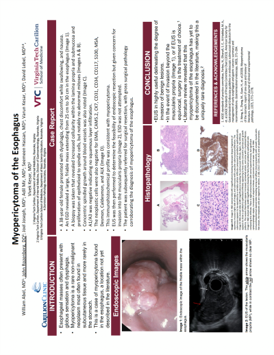
Introduction: Esophageal masses often present with symptoms of globus sensation, dysphagia, and associated weight loss. Benign lesions may also present with the same symptoms, making imaging and biopsy invaluable for a final diagnosis. Myopericytoma is a rare non-malignant neoplasm arising as a result of a PDGFRB mutation that is most commonly found in subcutaneous tissue. Histopathologic features include multilayered concentric growth around blood vessels and a well-circumscribed nodular appearance. We present a case of myopericytoma of the esophagus, a location not yet documented in the literature.
Case Description/Methods: Patient is a 38-year-old female with no significant past medical history who presented with symptoms of dysphagia, nausea, and chest pain with food intake. The symptoms were refractory to proton pump inhibitor (PPI) treatment as well as sucralfate. Esophagogastroduodenoscopy (EGD) examination revealed a large friable mass extending from 25 to 30 cm into the distal esophagus. Histopathology revealed increased cellularity within the lamina propria and submucosa with proliferation of epithelioid to spindled cells in the background stroma, but with no significant atypia or abnormal mitoses. There was concentric growth spindled cells around blood vessels. A1/A3 were negative within the stromal cells, showing no squamous infiltration. The neoplastic cells were also negative for EMA, CAM5.2, CK7, CD31, CD34, CD117, S100, MSA, Desmon, Caldesmon, and ALK. The immunoprofile and histopathology were consistent with myopericytoma. Further endoscopic evaluation proceeded with endoscopic ultrasound (EUS) to determine the feasibility of endoscopic resection but given concern for size and depth of invasion into the muscularis propria, the patient was referred for surgical removal of the mass.
Discussion: Myopericytomas are highly rare type of benign tumor, and if a surgical specimen is inadequate these may even be misdiagnosed as sarcomas. When combined with EUS, the surgical pathology specimen allowed for this patient to proceed directly to surgical treatment given involvement of the muscularis propria layer. Given benign nature of the tumor however, endoscopic resection with endoscopic submucosal dissection (ESD) is likely feasible under the right circumstances. This case highlights the importance of a multidisciplinary approach between surgeons and gastroenterologists. Furthermore, literature review reveals that myopericytoma of the esophagus has yet to be reported.
Disclosures:
William Abel, MD1, John Rozenberg, MD1, Joel Joseph, MD1, Adil Mir, MD1, Seemeen Hassan, MD1, Varun Kesar, MD1, David Lebel, MD2, Vivek Kesar, MD1. P0571 - Myopericytoma of the Esophagus: A New Location, ACG 2024 Annual Scientific Meeting Abstracts. Philadelphia, PA: American College of Gastroenterology.
1Carilion Clinic, Roanoke, VA; 2Dominion Pathology, Roanoke, VA
Introduction: Esophageal masses often present with symptoms of globus sensation, dysphagia, and associated weight loss. Benign lesions may also present with the same symptoms, making imaging and biopsy invaluable for a final diagnosis. Myopericytoma is a rare non-malignant neoplasm arising as a result of a PDGFRB mutation that is most commonly found in subcutaneous tissue. Histopathologic features include multilayered concentric growth around blood vessels and a well-circumscribed nodular appearance. We present a case of myopericytoma of the esophagus, a location not yet documented in the literature.
Case Description/Methods: Patient is a 38-year-old female with no significant past medical history who presented with symptoms of dysphagia, nausea, and chest pain with food intake. The symptoms were refractory to proton pump inhibitor (PPI) treatment as well as sucralfate. Esophagogastroduodenoscopy (EGD) examination revealed a large friable mass extending from 25 to 30 cm into the distal esophagus. Histopathology revealed increased cellularity within the lamina propria and submucosa with proliferation of epithelioid to spindled cells in the background stroma, but with no significant atypia or abnormal mitoses. There was concentric growth spindled cells around blood vessels. A1/A3 were negative within the stromal cells, showing no squamous infiltration. The neoplastic cells were also negative for EMA, CAM5.2, CK7, CD31, CD34, CD117, S100, MSA, Desmon, Caldesmon, and ALK. The immunoprofile and histopathology were consistent with myopericytoma. Further endoscopic evaluation proceeded with endoscopic ultrasound (EUS) to determine the feasibility of endoscopic resection but given concern for size and depth of invasion into the muscularis propria, the patient was referred for surgical removal of the mass.
Discussion: Myopericytomas are highly rare type of benign tumor, and if a surgical specimen is inadequate these may even be misdiagnosed as sarcomas. When combined with EUS, the surgical pathology specimen allowed for this patient to proceed directly to surgical treatment given involvement of the muscularis propria layer. Given benign nature of the tumor however, endoscopic resection with endoscopic submucosal dissection (ESD) is likely feasible under the right circumstances. This case highlights the importance of a multidisciplinary approach between surgeons and gastroenterologists. Furthermore, literature review reveals that myopericytoma of the esophagus has yet to be reported.
Disclosures:
William Abel indicated no relevant financial relationships.
John Rozenberg indicated no relevant financial relationships.
Joel Joseph indicated no relevant financial relationships.
Adil Mir indicated no relevant financial relationships.
Seemeen Hassan indicated no relevant financial relationships.
Varun Kesar indicated no relevant financial relationships.
David Lebel indicated no relevant financial relationships.
Vivek Kesar indicated no relevant financial relationships.
William Abel, MD1, John Rozenberg, MD1, Joel Joseph, MD1, Adil Mir, MD1, Seemeen Hassan, MD1, Varun Kesar, MD1, David Lebel, MD2, Vivek Kesar, MD1. P0571 - Myopericytoma of the Esophagus: A New Location, ACG 2024 Annual Scientific Meeting Abstracts. Philadelphia, PA: American College of Gastroenterology.
