Sunday Poster Session
Category: General Endoscopy
P0694 - Clinical Spectrum of Gastrointestinal Manifestations Related to Sevelamer Crystal Deposition at a Tertiary Care Hospital: A Case Series
Sunday, October 27, 2024
3:30 PM - 7:00 PM ET
Location: Exhibit Hall E

Has Audio
- AM
Aun Muhammad, MD
University of Mississippi Medical Center
Jackson, MS
Presenting Author(s)
Aun Muhammad, MD1, Zehra Naseem, MD2, John G. Ingram, DO1, Saleha Aziz, MD1, William P. Daley, MD1, Hamid Ullah, MBBS1, Mohamed Hassanein, MD1, Joydeep Chakraborty, MD1
1University of Mississippi Medical Center, Jackson, MS; 2Cleveland Clinic, Cleveland, OH
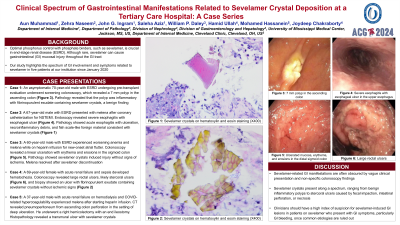
Introduction: Optimal phosphorus control with phosphate binders, such as sevelamer, is crucial in end-stage renal disease (ESRD). Although rare, sevelamer can cause gastrointestinal (GI) mucosal injury throughout the GI tract. Our study highlights the spectrum of GI involvement and symptoms related to sevelamer in five patients at our institution since January 2020.
Case Description/Methods: Case 1: An asymptomatic 70-year-old male with ESRD undergoing pre-transplant evaluation underwent screening colonoscopy, which revealed a 7 mm polyp in the ascending colon. Pathology revealed that the polyp was inflammatory with fibrinopurulent exudate containing sevelamer crystals, a benign finding.
Case 2: A 67-year-old male with ESRD presented with melena after coronary catheterization for NSTEMI. Endoscopy revealed severe esophagitis with esophageal ulcer. Pathology showed acute esophagitis with ulceration, necroinflammatory debris, and fish scale-like foreign material consistent with sevelamer crystals.
Case 3: A 60-year-old male with ESRD experienced worsening anemia and melena while on heparin infusion for new-onset atrial flutter. Colonoscopy revealed a linear ulceration with erythema and erosions in the sigmoid colon. Pathology showed sevelamer crystals induced injury without signs of ischemia. Melena resolved after sevelamer discontinuation.
Case 4: A 69-year-old female with acute renal failure and sepsis developed hematochezia. Colonoscopy revealed large rectal ulcers, likely stercoral ulcers, and biopsy showed an ulcer with fibrinopurulent exudate containing sevelamer crystals without ischemic signs.
Case 5: A 37-year-old male with acute renal failure on hemodialysis and COVID-related hypercoagulability experienced melena after starting heparin infusion. CT revealed pneumoperitoneum from ascending colon perforation in the setting of deep ulceration. He underwent a right hemicolectomy with an end ileostomy. Histopathology revealed a transmural ulcer with sevelamer crystals.
Discussion: Sevelamer-related GI manifestations are often obscured by vague clinical presentation and non-specific colonoscopy findings. Sevelamer crystals present along a spectrum, ranging from benign inflammatory polyps to stercoral ulcers caused by fecal impaction, intestinal perforation, or necrosis. Clinicians should have a high index of suspicion for sevelamer-induced GI lesions in patients on sevelamer who present with GI symptoms, particularly GI bleeding, once common etiologies are ruled out.
Disclosures:
Aun Muhammad, MD1, Zehra Naseem, MD2, John G. Ingram, DO1, Saleha Aziz, MD1, William P. Daley, MD1, Hamid Ullah, MBBS1, Mohamed Hassanein, MD1, Joydeep Chakraborty, MD1. P0694 - Clinical Spectrum of Gastrointestinal Manifestations Related to Sevelamer Crystal Deposition at a Tertiary Care Hospital: A Case Series, ACG 2024 Annual Scientific Meeting Abstracts. Philadelphia, PA: American College of Gastroenterology.
1University of Mississippi Medical Center, Jackson, MS; 2Cleveland Clinic, Cleveland, OH
Introduction: Optimal phosphorus control with phosphate binders, such as sevelamer, is crucial in end-stage renal disease (ESRD). Although rare, sevelamer can cause gastrointestinal (GI) mucosal injury throughout the GI tract. Our study highlights the spectrum of GI involvement and symptoms related to sevelamer in five patients at our institution since January 2020.
Case Description/Methods: Case 1: An asymptomatic 70-year-old male with ESRD undergoing pre-transplant evaluation underwent screening colonoscopy, which revealed a 7 mm polyp in the ascending colon. Pathology revealed that the polyp was inflammatory with fibrinopurulent exudate containing sevelamer crystals, a benign finding.
Case 2: A 67-year-old male with ESRD presented with melena after coronary catheterization for NSTEMI. Endoscopy revealed severe esophagitis with esophageal ulcer. Pathology showed acute esophagitis with ulceration, necroinflammatory debris, and fish scale-like foreign material consistent with sevelamer crystals.
Case 3: A 60-year-old male with ESRD experienced worsening anemia and melena while on heparin infusion for new-onset atrial flutter. Colonoscopy revealed a linear ulceration with erythema and erosions in the sigmoid colon. Pathology showed sevelamer crystals induced injury without signs of ischemia. Melena resolved after sevelamer discontinuation.
Case 4: A 69-year-old female with acute renal failure and sepsis developed hematochezia. Colonoscopy revealed large rectal ulcers, likely stercoral ulcers, and biopsy showed an ulcer with fibrinopurulent exudate containing sevelamer crystals without ischemic signs.
Case 5: A 37-year-old male with acute renal failure on hemodialysis and COVID-related hypercoagulability experienced melena after starting heparin infusion. CT revealed pneumoperitoneum from ascending colon perforation in the setting of deep ulceration. He underwent a right hemicolectomy with an end ileostomy. Histopathology revealed a transmural ulcer with sevelamer crystals.
Discussion: Sevelamer-related GI manifestations are often obscured by vague clinical presentation and non-specific colonoscopy findings. Sevelamer crystals present along a spectrum, ranging from benign inflammatory polyps to stercoral ulcers caused by fecal impaction, intestinal perforation, or necrosis. Clinicians should have a high index of suspicion for sevelamer-induced GI lesions in patients on sevelamer who present with GI symptoms, particularly GI bleeding, once common etiologies are ruled out.
Disclosures:
Aun Muhammad indicated no relevant financial relationships.
Zehra Naseem indicated no relevant financial relationships.
John G. Ingram indicated no relevant financial relationships.
Saleha Aziz indicated no relevant financial relationships.
William P. Daley indicated no relevant financial relationships.
Hamid Ullah indicated no relevant financial relationships.
Mohamed Hassanein indicated no relevant financial relationships.
Joydeep Chakraborty indicated no relevant financial relationships.
Aun Muhammad, MD1, Zehra Naseem, MD2, John G. Ingram, DO1, Saleha Aziz, MD1, William P. Daley, MD1, Hamid Ullah, MBBS1, Mohamed Hassanein, MD1, Joydeep Chakraborty, MD1. P0694 - Clinical Spectrum of Gastrointestinal Manifestations Related to Sevelamer Crystal Deposition at a Tertiary Care Hospital: A Case Series, ACG 2024 Annual Scientific Meeting Abstracts. Philadelphia, PA: American College of Gastroenterology.
