Sunday Poster Session
Category: General Endoscopy
P0703 - Battling the Burn: A Case of Endoscopically Diagnosed Severe Caustic Injury
Sunday, October 27, 2024
3:30 PM - 7:00 PM ET
Location: Exhibit Hall E

Has Audio

Pratiksha Moliya, MD
Jamaica Hospital Medical Center
Edison, NJ
Presenting Author(s)
Pratiksha Moliya, MD1, Ibrahim Nabous, MD2, Hasan Al-Obaidi, MD3, Srikaran Bojja, MD4, Sophia Jagroop, MD5
1Jamaica Hospital Medical Center, Edison, NJ; 2Jamaica Hospital Medical Center, Elmont, NY; 3Jamaica Hospital Medical Center, Briarwood, NY; 4BronxCare Health System, Bronx, NY; 5Jamaica Hospital Medical Center, Queens, NY
Introduction: Caustic injuries account for about 10% of poisoning cases with an 8% mortality rate, highlighting their severity. These injuries challenge gastroenterologists, with endoscopy (EGD) playing a crucial role. Here, we present a case of an endoscopically diagnosed caustic injury.
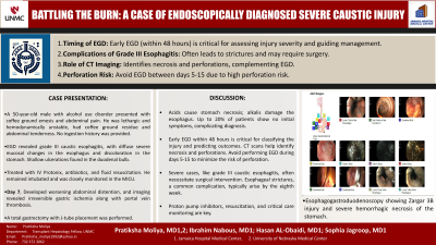
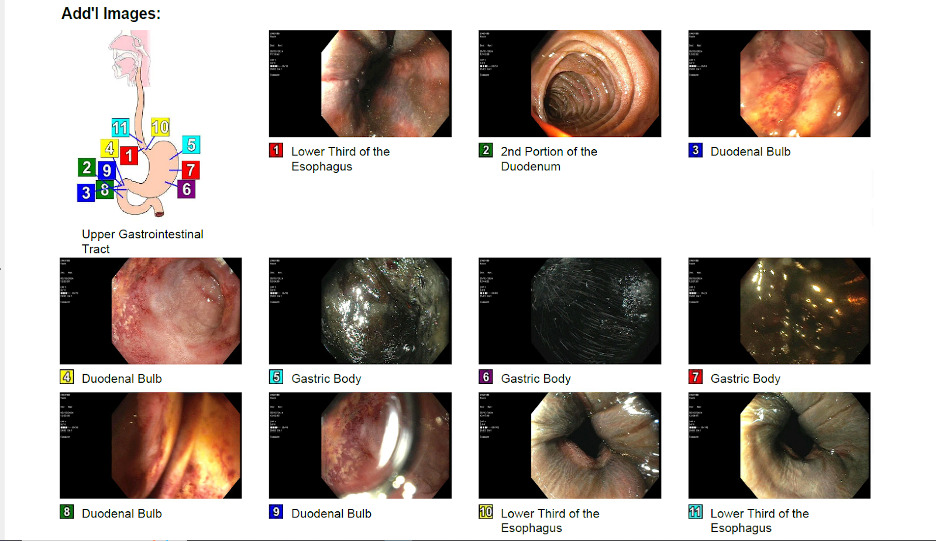
Case Description/Methods: A 50-year-old male with a history of alcohol use presented with coffee ground emesis and abdominal pain. He did not provide ingestion history. Upon arrival, he was lethargic, tachycardic, hypotensive, and hypoxic. Examination revealed small amounts of coffee ground residue in his throat and generalized abdominal tenderness. He required vasopressors and was intubated for airway protection. Initial labs showed mild neutrophilic leukocytosis and transaminitis, but surprisingly no anemia. A CT abdomen showed a thickened wall and distended stomach. EGD revealed grade III caustic esophagitis throughout the esophagus, with diffuse severe mucosal changes and discoloration in the stomach. Shallow ulcerations were noted in the duodenal bulb. He was admitted to the MICU and treated with IV Protonix, antibiotics, and fluid resuscitation. After a week, he developed worsening abdominal distension. Imaging showed irreversible stomach ischemia with portal vein thrombosis, leading to a total gastrectomy and J-tube placement.
Discussion: Caustic ingestions in adults are severe and often intentional, with acids causing coagulation necrosis in the stomach and alkalis causing liquefaction necrosis in the esophagus. About 20% of patients show no initial oropharyngeal symptoms and may not provide a clear ingestion history, as in our patient. Protocols recommend early EGD within 48 hours, as classification and severity of the injury predict outcomes, with grade IIIb patients experiencing longer hospital stays and more complications than grade IIIa. In our case, EGD within 8 hours of arrival was crucial for diagnosing and assessing the injury. CT scans identify necrosis and potential perforations. EGD is avoided from days 5-15 due to perforation risks. Management includes proton pump inhibitors, resuscitation, and critical care monitoring. Grade III caustic esophagitis frequently requires surgical intervention. Esophageal strictures, common in severe cases, usually develop by the eighth week. Our case underscores the importance of early radiological and endoscopic evaluations, as history and symptoms might be unreliable. Early evaluations are vital for assessing prognosis and minimizing complications in caustic injuries.

Disclosures:
Pratiksha Moliya, MD1, Ibrahim Nabous, MD2, Hasan Al-Obaidi, MD3, Srikaran Bojja, MD4, Sophia Jagroop, MD5. P0703 - Battling the Burn: A Case of Endoscopically Diagnosed Severe Caustic Injury, ACG 2024 Annual Scientific Meeting Abstracts. Philadelphia, PA: American College of Gastroenterology.
1Jamaica Hospital Medical Center, Edison, NJ; 2Jamaica Hospital Medical Center, Elmont, NY; 3Jamaica Hospital Medical Center, Briarwood, NY; 4BronxCare Health System, Bronx, NY; 5Jamaica Hospital Medical Center, Queens, NY
Introduction: Caustic injuries account for about 10% of poisoning cases with an 8% mortality rate, highlighting their severity. These injuries challenge gastroenterologists, with endoscopy (EGD) playing a crucial role. Here, we present a case of an endoscopically diagnosed caustic injury.
Case Description/Methods: A 50-year-old male with a history of alcohol use presented with coffee ground emesis and abdominal pain. He did not provide ingestion history. Upon arrival, he was lethargic, tachycardic, hypotensive, and hypoxic. Examination revealed small amounts of coffee ground residue in his throat and generalized abdominal tenderness. He required vasopressors and was intubated for airway protection. Initial labs showed mild neutrophilic leukocytosis and transaminitis, but surprisingly no anemia. A CT abdomen showed a thickened wall and distended stomach. EGD revealed grade III caustic esophagitis throughout the esophagus, with diffuse severe mucosal changes and discoloration in the stomach. Shallow ulcerations were noted in the duodenal bulb. He was admitted to the MICU and treated with IV Protonix, antibiotics, and fluid resuscitation. After a week, he developed worsening abdominal distension. Imaging showed irreversible stomach ischemia with portal vein thrombosis, leading to a total gastrectomy and J-tube placement.
Discussion: Caustic ingestions in adults are severe and often intentional, with acids causing coagulation necrosis in the stomach and alkalis causing liquefaction necrosis in the esophagus. About 20% of patients show no initial oropharyngeal symptoms and may not provide a clear ingestion history, as in our patient. Protocols recommend early EGD within 48 hours, as classification and severity of the injury predict outcomes, with grade IIIb patients experiencing longer hospital stays and more complications than grade IIIa. In our case, EGD within 8 hours of arrival was crucial for diagnosing and assessing the injury. CT scans identify necrosis and potential perforations. EGD is avoided from days 5-15 due to perforation risks. Management includes proton pump inhibitors, resuscitation, and critical care monitoring. Grade III caustic esophagitis frequently requires surgical intervention. Esophageal strictures, common in severe cases, usually develop by the eighth week. Our case underscores the importance of early radiological and endoscopic evaluations, as history and symptoms might be unreliable. Early evaluations are vital for assessing prognosis and minimizing complications in caustic injuries.

Figure: Esophagogastroduodenoscopy showing Zargar 3B injury and severe hemorrhagic necrosis of the stomach.
Disclosures:
Pratiksha Moliya indicated no relevant financial relationships.
Ibrahim Nabous indicated no relevant financial relationships.
Hasan Al-Obaidi indicated no relevant financial relationships.
Srikaran Bojja indicated no relevant financial relationships.
Sophia Jagroop indicated no relevant financial relationships.
Pratiksha Moliya, MD1, Ibrahim Nabous, MD2, Hasan Al-Obaidi, MD3, Srikaran Bojja, MD4, Sophia Jagroop, MD5. P0703 - Battling the Burn: A Case of Endoscopically Diagnosed Severe Caustic Injury, ACG 2024 Annual Scientific Meeting Abstracts. Philadelphia, PA: American College of Gastroenterology.
