Sunday Poster Session
Category: General Endoscopy
P0706 - PEG Tube Gone Wrong! Percutaneous Endoscopic Gastrostomy Transversing the Transverse Colon
Sunday, October 27, 2024
3:30 PM - 7:00 PM ET
Location: Exhibit Hall E

Has Audio
- ST
Shunsa Tarar, DO
Albany Medical Center
Albany, NY
Presenting Author(s)
Shunsa Tarar, DO1, Ahmad Abulawi, MD2, James Puleo, MD1
1Albany Medical Center, Albany, NY; 2Albany Medical College, Albany, NY
Introduction: Percutaneous endoscopic gastrostomy (PEG) placement is a minimally invasive mechanism to sustain nutrition in patients requiring long-term supplementation. Risks associated with this technique range from pneumoperitoneum to perforation and bleeding. We present a case of PEG placement transversing the transverse colon requiring surgical removal and placement of gastrostomy tube.
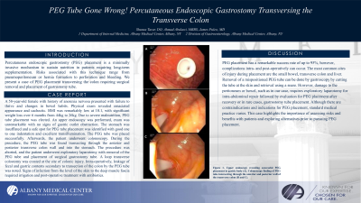
Case Description/Methods: A 58-year-old female with history of anorexia nervosa presented with failure to thrive and changes in bowel habits. Physical exam revealed emaciated appearance and cachectic. BMI was remarkably low at 10.1, with significant weight loss over 6 months from 44 kilo grams (kg) to 26 kg. PEG tube placement was elected and colonoscopy for evaluation of changes in bowel habits. An upper endoscopy was performed, exam was unremarkable. The stomach was insufflated and a safe spot for PEG tube placement was identified with good one to one indentation and excellent transillumination. The PEG tube was placed successfully. Afterwards, the patient underwent diagnostic colonoscopy for changes in bowel habits. During the procedure, the PEG tube was found in the transverse colon intersecting through the anterior and posterior transverse colon wall and into the stomach. The patient underwent exploratory laparotomy with removal of the PEG tube and placement of surgical gastrostomy tube at the same site. Due to the patient’s history of failure to thrive, a loop transverse colostomy was created at the site of colonic injury. Intra-operatively, leakage of fecal and gastric contents secondary to transection of the colon by the PEG tube was noted. Signs of infection from the level of the skin to the deep muscle fascia required irrigation and treatment with antibiotics.
Discussion: PEG placement has a remarkable success rate of 95%, however, complications intra- and post-operatively can occur. Removal of a mispositioned PEG tube can be done by gastroscopy by cutting the tube at the skin and retrieval using a snare. Subsequently, defects closure by clips. However, damage to the peritoneum or bowel, such as in our case, requires exploratory laparotomy for intra-abdominal repair followed by evaluation for PEG placement after recovery or in rare cases, gastrostomy tube placement.
Although there are contraindications and indications for PEG placement, standard medical practice varies. This case highlights the importance of assessing risks and benefits with patients and exploring alternatives prior to pursuing PEG placement.

Disclosures:
Shunsa Tarar, DO1, Ahmad Abulawi, MD2, James Puleo, MD1. P0706 - PEG Tube Gone Wrong! Percutaneous Endoscopic Gastrostomy Transversing the Transverse Colon, ACG 2024 Annual Scientific Meeting Abstracts. Philadelphia, PA: American College of Gastroenterology.
1Albany Medical Center, Albany, NY; 2Albany Medical College, Albany, NY
Introduction: Percutaneous endoscopic gastrostomy (PEG) placement is a minimally invasive mechanism to sustain nutrition in patients requiring long-term supplementation. Risks associated with this technique range from pneumoperitoneum to perforation and bleeding. We present a case of PEG placement transversing the transverse colon requiring surgical removal and placement of gastrostomy tube.
Case Description/Methods: A 58-year-old female with history of anorexia nervosa presented with failure to thrive and changes in bowel habits. Physical exam revealed emaciated appearance and cachectic. BMI was remarkably low at 10.1, with significant weight loss over 6 months from 44 kilo grams (kg) to 26 kg. PEG tube placement was elected and colonoscopy for evaluation of changes in bowel habits. An upper endoscopy was performed, exam was unremarkable. The stomach was insufflated and a safe spot for PEG tube placement was identified with good one to one indentation and excellent transillumination. The PEG tube was placed successfully. Afterwards, the patient underwent diagnostic colonoscopy for changes in bowel habits. During the procedure, the PEG tube was found in the transverse colon intersecting through the anterior and posterior transverse colon wall and into the stomach. The patient underwent exploratory laparotomy with removal of the PEG tube and placement of surgical gastrostomy tube at the same site. Due to the patient’s history of failure to thrive, a loop transverse colostomy was created at the site of colonic injury. Intra-operatively, leakage of fecal and gastric contents secondary to transection of the colon by the PEG tube was noted. Signs of infection from the level of the skin to the deep muscle fascia required irrigation and treatment with antibiotics.
Discussion: PEG placement has a remarkable success rate of 95%, however, complications intra- and post-operatively can occur. Removal of a mispositioned PEG tube can be done by gastroscopy by cutting the tube at the skin and retrieval using a snare. Subsequently, defects closure by clips. However, damage to the peritoneum or bowel, such as in our case, requires exploratory laparotomy for intra-abdominal repair followed by evaluation for PEG placement after recovery or in rare cases, gastrostomy tube placement.
Although there are contraindications and indications for PEG placement, standard medical practice varies. This case highlights the importance of assessing risks and benefits with patients and exploring alternatives prior to pursuing PEG placement.

Figure: Figure 1: Upper endoscopy revealing successful PEG placement in gastric body (A). Colonoscopy findings of PEG tube intersecting through the anterior and posterior wall of the transverse colon (B and C).
Disclosures:
Shunsa Tarar indicated no relevant financial relationships.
Ahmad Abulawi indicated no relevant financial relationships.
James Puleo indicated no relevant financial relationships.
Shunsa Tarar, DO1, Ahmad Abulawi, MD2, James Puleo, MD1. P0706 - PEG Tube Gone Wrong! Percutaneous Endoscopic Gastrostomy Transversing the Transverse Colon, ACG 2024 Annual Scientific Meeting Abstracts. Philadelphia, PA: American College of Gastroenterology.
