Sunday Poster Session
Category: GI Bleeding
P0776 - Gastrointestinal Stromal Tumors Presenting as Recurrent Gastrointestinal Bleeding
Sunday, October 27, 2024
3:30 PM - 7:00 PM ET
Location: Exhibit Hall E

Has Audio
- NM
Nadia Mishal, MD
Lewis Gale Medical Center
Salem, VA
Presenting Author(s)
Safia Afaq, MD1, Taimur Khan, MD2, Nadia Mishal, MD1, Chuan L. Miao, MD, MPH, MSc, FACG1
1Lewis Gale Medical Center, Salem, VA; 2Post Medical School, Salem, VA
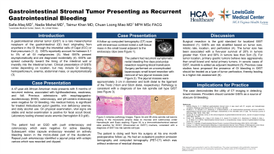
Introduction: A gastrointestinal stromal tumor (GIST) is a rare malignant mesenchymal neoplasm of the gastrointestinal (GI) tract. The clinical presentation of GIST tumors varies depending on location of disease and includes GI bleeding, hemoperitoneum, anemia, abdominal mass, or absence of symptoms. Most GISTs present asymptomatically and are diagnosed incidentally on imaging or surgically. This case involves recurrent GI bleeding from two jejunal GIST tumors.
Case Description/Methods: A 47-year-old African American male presents with five months of recurrent melena and symptomatic anemia. His prior work up includes esophagogastroduodenoscopy (EGD), colonoscopy, and pill camera endoscopy which were negative for a bleeding source. His medical history is significant for treated Helicobacter pylori gastritis and alcohol dependence. On examination, vital signs are stable, rectal examination is positive for fecal occult blood, and labs show a hemoglobin 8.9 g/dl. EGD with push enteroscopy and colonoscopy were negative for active bleeding, though small capsule endoscopy found an actively bleeding lesion in the mid-to-distal part of the duodenum. Repeat push enteroscopy found a jejunal polyp with ectopic varices which was resected and clipped. A computed tomography (CT) scan with intravenous contrast noted a soft tissue mass in the small bowel near the endoscopy clips. Surgery performed a laparoscopic small bowel resection with side-to-side anastomosis and removed two jejunal masses. The pathology diagnosed low risk spindle cell type GIST tumors. A positron emission tomography and computed tomography (PET-CT) scan for tumor staging was negative for residual disease.
Discussion: The events of this case occurred over a span of five months with difficulty in locating the source. This is frequently the case in small intestinal GI bleeding. If a source of bleeding is not found on endoscopy, the next recommended step is for CT imaging. Surgical resection is the gold standard treatment for localized GIST. In more severe cases of GIST, imatinib is added as adjuvant treatment. In this case, the tumor was a low risk spindle cell type and no residual disease was found on PET-CT, so imatinib was not initiated. Low risk GIST tumors are reported to have recurrence up to 20 years after surgical resection. Yet, previous case studies have proposed the presence of GI bleeding in GIST tumors should be treated as a type of tumor perforation and have a higher risk.

Disclosures:
Safia Afaq, MD1, Taimur Khan, MD2, Nadia Mishal, MD1, Chuan L. Miao, MD, MPH, MSc, FACG1. P0776 - Gastrointestinal Stromal Tumors Presenting as Recurrent Gastrointestinal Bleeding, ACG 2024 Annual Scientific Meeting Abstracts. Philadelphia, PA: American College of Gastroenterology.
1Lewis Gale Medical Center, Salem, VA; 2Post Medical School, Salem, VA
Introduction: A gastrointestinal stromal tumor (GIST) is a rare malignant mesenchymal neoplasm of the gastrointestinal (GI) tract. The clinical presentation of GIST tumors varies depending on location of disease and includes GI bleeding, hemoperitoneum, anemia, abdominal mass, or absence of symptoms. Most GISTs present asymptomatically and are diagnosed incidentally on imaging or surgically. This case involves recurrent GI bleeding from two jejunal GIST tumors.
Case Description/Methods: A 47-year-old African American male presents with five months of recurrent melena and symptomatic anemia. His prior work up includes esophagogastroduodenoscopy (EGD), colonoscopy, and pill camera endoscopy which were negative for a bleeding source. His medical history is significant for treated Helicobacter pylori gastritis and alcohol dependence. On examination, vital signs are stable, rectal examination is positive for fecal occult blood, and labs show a hemoglobin 8.9 g/dl. EGD with push enteroscopy and colonoscopy were negative for active bleeding, though small capsule endoscopy found an actively bleeding lesion in the mid-to-distal part of the duodenum. Repeat push enteroscopy found a jejunal polyp with ectopic varices which was resected and clipped. A computed tomography (CT) scan with intravenous contrast noted a soft tissue mass in the small bowel near the endoscopy clips. Surgery performed a laparoscopic small bowel resection with side-to-side anastomosis and removed two jejunal masses. The pathology diagnosed low risk spindle cell type GIST tumors. A positron emission tomography and computed tomography (PET-CT) scan for tumor staging was negative for residual disease.
Discussion: The events of this case occurred over a span of five months with difficulty in locating the source. This is frequently the case in small intestinal GI bleeding. If a source of bleeding is not found on endoscopy, the next recommended step is for CT imaging. Surgical resection is the gold standard treatment for localized GIST. In more severe cases of GIST, imatinib is added as adjuvant treatment. In this case, the tumor was a low risk spindle cell type and no residual disease was found on PET-CT, so imatinib was not initiated. Low risk GIST tumors are reported to have recurrence up to 20 years after surgical resection. Yet, previous case studies have proposed the presence of GI bleeding in GIST tumors should be treated as a type of tumor perforation and have a higher risk.

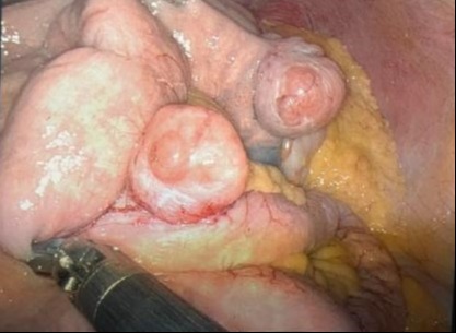
Figure: Figure shows the operative findings significant for two jejunal masses, one about 10 cm distal to the ligament of Treitz and the other about 40 cm distal from 1st mass, approximately 3 cm in diameter each.
Disclosures:
Safia Afaq indicated no relevant financial relationships.
Taimur Khan indicated no relevant financial relationships.
Nadia Mishal indicated no relevant financial relationships.
Chuan Miao indicated no relevant financial relationships.
Safia Afaq, MD1, Taimur Khan, MD2, Nadia Mishal, MD1, Chuan L. Miao, MD, MPH, MSc, FACG1. P0776 - Gastrointestinal Stromal Tumors Presenting as Recurrent Gastrointestinal Bleeding, ACG 2024 Annual Scientific Meeting Abstracts. Philadelphia, PA: American College of Gastroenterology.

