Sunday Poster Session
Category: GI Bleeding
P0805 - Jejunal Ectopic Varices: Obscure Bleeding Culprit After Sleeve Gastrectomy Found With Video Capsule Endoscopy
Sunday, October 27, 2024
3:30 PM - 7:00 PM ET
Location: Exhibit Hall E

Has Audio

Swati Mahapatra, DO
TTUHSC
El Paso, TX
Presenting Author(s)
Swati Mahapatra, DO1, Monica Botros, MD1, Shivangini Duggal, MD2, Jesus A. Guzman, MD1, Ricardo Badillo, MD2
1Texas Tech University Health Sciences Center, El Paso, TX; 2TTUHSC, El Paso, TX
Introduction: Ectopic varices are dilated portosystemic collateral veins that are usually located in a different region other than the gastroesophageal area and are associated with a higher risk of rebleeding and higher mortality rates. They account for up to 5% of variceal bleeding and can present with signs of both upper or lower gastrointestinal (GI) bleeding depending on the location. The duodenum is the most frequent site for small bowel varices followed by jejunum and ileum; other sites include colon and peritoneum. Portal hypertension (PH) is the most common etiology. Management is challenging due to difficulty in localization and lack of clear guidelines for treatment.
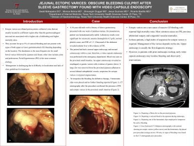
Case Description/Methods: A 54-year-old male with a history of sleeve gastrectomy, presented with one week of painless melena. On presentation, patient was hemodynamically stable. Laboratory results were significant for microcytic anemia (hemoglobin 6.5 g/dl), normal platelet count, and INR of 1.3. Ultrasound of the abdomen revealed nodular liver with evidence of PH. The patient had had a normal upper endoscopy and normal colonoscopy within a year; therefore, a video capsule endoscopy was performed in the emergency department. Blood was seen in the proximal small intestine. An upper enteroscopy revealed no esophageal or gastric varices with evidence of gastric sleeve. A large clot was removed from the proximal jejunum adherent to several dilated subepithelial vessels, suspicious for ectopic varices vs atypical angioectasias. To temporize the bleeding for definitive therapy, 3 hemostatic clips were placed and no further bleeding reported (Figure 1). CT enterography after the procedure confirmed the presence of PH and ectopic varices in the proximal small intestine (Figure 2).
Discussion: Ectopic varices are a rare cause of massive GI bleeding with reported high mortality rates. Most common causes are PH, previous abdominal surgery and congenital vascular anomalies. In these patients, a high index of suspicion for ectopic varices is required. Management of the varices depends on the site. Upper endoscopy is usually the first diagnostic strategy. However, in patients with prior endoscopic workup, early video capsule endoscopy may localize bleeding and direct early interventions.

Disclosures:
Swati Mahapatra, DO1, Monica Botros, MD1, Shivangini Duggal, MD2, Jesus A. Guzman, MD1, Ricardo Badillo, MD2. P0805 - Jejunal Ectopic Varices: Obscure Bleeding Culprit After Sleeve Gastrectomy Found With Video Capsule Endoscopy, ACG 2024 Annual Scientific Meeting Abstracts. Philadelphia, PA: American College of Gastroenterology.
1Texas Tech University Health Sciences Center, El Paso, TX; 2TTUHSC, El Paso, TX
Introduction: Ectopic varices are dilated portosystemic collateral veins that are usually located in a different region other than the gastroesophageal area and are associated with a higher risk of rebleeding and higher mortality rates. They account for up to 5% of variceal bleeding and can present with signs of both upper or lower gastrointestinal (GI) bleeding depending on the location. The duodenum is the most frequent site for small bowel varices followed by jejunum and ileum; other sites include colon and peritoneum. Portal hypertension (PH) is the most common etiology. Management is challenging due to difficulty in localization and lack of clear guidelines for treatment.
Case Description/Methods: A 54-year-old male with a history of sleeve gastrectomy, presented with one week of painless melena. On presentation, patient was hemodynamically stable. Laboratory results were significant for microcytic anemia (hemoglobin 6.5 g/dl), normal platelet count, and INR of 1.3. Ultrasound of the abdomen revealed nodular liver with evidence of PH. The patient had had a normal upper endoscopy and normal colonoscopy within a year; therefore, a video capsule endoscopy was performed in the emergency department. Blood was seen in the proximal small intestine. An upper enteroscopy revealed no esophageal or gastric varices with evidence of gastric sleeve. A large clot was removed from the proximal jejunum adherent to several dilated subepithelial vessels, suspicious for ectopic varices vs atypical angioectasias. To temporize the bleeding for definitive therapy, 3 hemostatic clips were placed and no further bleeding reported (Figure 1). CT enterography after the procedure confirmed the presence of PH and ectopic varices in the proximal small intestine (Figure 2).
Discussion: Ectopic varices are a rare cause of massive GI bleeding with reported high mortality rates. Most common causes are PH, previous abdominal surgery and congenital vascular anomalies. In these patients, a high index of suspicion for ectopic varices is required. Management of the varices depends on the site. Upper endoscopy is usually the first diagnostic strategy. However, in patients with prior endoscopic workup, early video capsule endoscopy may localize bleeding and direct early interventions.

Figure: Figure 1: Upper enteroscopy findings pre and post hemostatic clips
Figure 2: Yellow arrow pointing to Jejunal varices, orange arrow pointing to the hemostatic clips
Figure 2: Yellow arrow pointing to Jejunal varices, orange arrow pointing to the hemostatic clips
Disclosures:
Swati Mahapatra indicated no relevant financial relationships.
Monica Botros indicated no relevant financial relationships.
Shivangini Duggal indicated no relevant financial relationships.
Jesus Guzman indicated no relevant financial relationships.
Ricardo Badillo indicated no relevant financial relationships.
Swati Mahapatra, DO1, Monica Botros, MD1, Shivangini Duggal, MD2, Jesus A. Guzman, MD1, Ricardo Badillo, MD2. P0805 - Jejunal Ectopic Varices: Obscure Bleeding Culprit After Sleeve Gastrectomy Found With Video Capsule Endoscopy, ACG 2024 Annual Scientific Meeting Abstracts. Philadelphia, PA: American College of Gastroenterology.
