Sunday Poster Session
Category: GI Bleeding
P0807 - Pyogenic Granuloma: A Rare Cause of Obscure Occult GI Bleeding
Sunday, October 27, 2024
3:30 PM - 7:00 PM ET
Location: Exhibit Hall E

Has Audio
- MS
Michael Siegel, DO
University of Illinois
Chicago, IL
Presenting Author(s)
Michael Siegel, DO1, Omar T. Ahmed, MD2, Saul E. Turcios Escobar, MD3, Grace Guzman, MD1, Wadih Chacra, MD3
1University of Illinois, Chicago, IL; 2University of Illinois at Chicago, Chicago, IL; 3University of Illinois College of Medicine, Chicago, IL
Introduction: A pyogenic granuloma (PG) is a polypoid lobular capillary hemangioma occurring on the skin and mucosal surfaces. Given their vascularity, they have a high propensity to bleed and can be a rare cause of severe anemia. To our knowledge, there are very few cases of PG occurring in other parts of the GI tract. We present a case of a pyogenic granuloma of the terminal ileum causing obscure occult gastrointestinal bleeding in a cirrhotic patient.
Case Description/Methods: A middle-aged female with decompensated MASH cirrhosis and portal hypertension complicated with a non-occlusive portal vein thrombus, not on anticoagulation, who presents to the ED with severe iron deficiency anemia hemoglobin of 5.9gm/dL from a baseline hemoglobin of 8 gm/dL, iron saturation 4% and ferritin 9 ng/ml despite iron supplementation. She denied any overt signs of bleeding including melena, hematochezia, hematemesis, or vaginal bleeding.
A repeat EGD was performed, which showed grade 1 esophageal varices with no stigmata of recent bleeding with portal hypertensive gastropathy. Subsequently, a video capsule endoscopy was done and showed small amounts of blood in the ileum without obvious source, and blood-tinged colon content. A colonoscopy with specks of heme seen in the entire colon. A careful examination of the terminal ileum showed one sessile, ulcerated 7mm polyp, about 10cm from the ileocecal valve (Fig A). Biopsies of the lesion showed focal acute ulcer and granulation tissue without evidence of malignancy. A repeat colonoscopy was performed for complete resection of the polyp done with a cold snare. Pathology showed a submucosal lobulated capillary hemangioma within villiform ileal mucosa consistent with ulcerated pyogenic granuloma (Fig B-C). The patient has had routine follow up since resection of the PG with stable hemoglobin levels and normal iron studies. The patient was started on apixaban for management of portal vein thrombosis without complications.
Discussion: Pyogenic granuloma is a rare benign vascular lesion of the GI tract from the oral cavity to the anus. Some cases are incidentally found during screening colonoscopies, other cases present with overt or occult GI bleeding leading to severe anemia. Very few cases of small bowel PG are reported in the literature as indicated a recent case series from 2020 with only 35 reported cases. The treatment is usually endoscopic resection with a snare polypectomy for smaller lesions while larger lesions may require a surgical intervention.

Disclosures:
Michael Siegel, DO1, Omar T. Ahmed, MD2, Saul E. Turcios Escobar, MD3, Grace Guzman, MD1, Wadih Chacra, MD3. P0807 - Pyogenic Granuloma: A Rare Cause of Obscure Occult GI Bleeding, ACG 2024 Annual Scientific Meeting Abstracts. Philadelphia, PA: American College of Gastroenterology.
1University of Illinois, Chicago, IL; 2University of Illinois at Chicago, Chicago, IL; 3University of Illinois College of Medicine, Chicago, IL
Introduction: A pyogenic granuloma (PG) is a polypoid lobular capillary hemangioma occurring on the skin and mucosal surfaces. Given their vascularity, they have a high propensity to bleed and can be a rare cause of severe anemia. To our knowledge, there are very few cases of PG occurring in other parts of the GI tract. We present a case of a pyogenic granuloma of the terminal ileum causing obscure occult gastrointestinal bleeding in a cirrhotic patient.
Case Description/Methods: A middle-aged female with decompensated MASH cirrhosis and portal hypertension complicated with a non-occlusive portal vein thrombus, not on anticoagulation, who presents to the ED with severe iron deficiency anemia hemoglobin of 5.9gm/dL from a baseline hemoglobin of 8 gm/dL, iron saturation 4% and ferritin 9 ng/ml despite iron supplementation. She denied any overt signs of bleeding including melena, hematochezia, hematemesis, or vaginal bleeding.
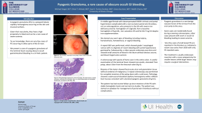
A repeat EGD was performed, which showed grade 1 esophageal varices with no stigmata of recent bleeding with portal hypertensive gastropathy. Subsequently, a video capsule endoscopy was done and showed small amounts of blood in the ileum without obvious source, and blood-tinged colon content. A colonoscopy with specks of heme seen in the entire colon. A careful examination of the terminal ileum showed one sessile, ulcerated 7mm polyp, about 10cm from the ileocecal valve (Fig A). Biopsies of the lesion showed focal acute ulcer and granulation tissue without evidence of malignancy. A repeat colonoscopy was performed for complete resection of the polyp done with a cold snare. Pathology showed a submucosal lobulated capillary hemangioma within villiform ileal mucosa consistent with ulcerated pyogenic granuloma (Fig B-C). The patient has had routine follow up since resection of the PG with stable hemoglobin levels and normal iron studies. The patient was started on apixaban for management of portal vein thrombosis without complications.
Discussion: Pyogenic granuloma is a rare benign vascular lesion of the GI tract from the oral cavity to the anus. Some cases are incidentally found during screening colonoscopies, other cases present with overt or occult GI bleeding leading to severe anemia. Very few cases of small bowel PG are reported in the literature as indicated a recent case series from 2020 with only 35 reported cases. The treatment is usually endoscopic resection with a snare polypectomy for smaller lesions while larger lesions may require a surgical intervention.

Figure: A: One sessile, ulcerated 7mm polyp, about 10cm from the ileocecal valve.
B: Histopathology examination of ileal polyp showing ulcerated pyogenic granuloma within villiform ileal mucosa
C: High power magnification, blood vessels are engorged with red blood cells.
B: Histopathology examination of ileal polyp showing ulcerated pyogenic granuloma within villiform ileal mucosa
C: High power magnification, blood vessels are engorged with red blood cells.
Disclosures:
Michael Siegel indicated no relevant financial relationships.
Omar Ahmed indicated no relevant financial relationships.
Saul Turcios Escobar indicated no relevant financial relationships.
Grace Guzman indicated no relevant financial relationships.
Wadih Chacra indicated no relevant financial relationships.
Michael Siegel, DO1, Omar T. Ahmed, MD2, Saul E. Turcios Escobar, MD3, Grace Guzman, MD1, Wadih Chacra, MD3. P0807 - Pyogenic Granuloma: A Rare Cause of Obscure Occult GI Bleeding, ACG 2024 Annual Scientific Meeting Abstracts. Philadelphia, PA: American College of Gastroenterology.
