Sunday Poster Session
Category: GI Bleeding
P0812 - Gangliocytic Paraganglioma: Ampullary Neoplasm as a Rare Cause of Upper Gastrointestinal Bleeding
Sunday, October 27, 2024
3:30 PM - 7:00 PM ET
Location: Exhibit Hall E

Has Audio

Joseph Granade, MD
Medical University of South Carolina
Charleston, SC
Presenting Author(s)
Joseph Granade, MD, Robert A. Moran, MD
Medical University of South Carolina, Charleston, SC
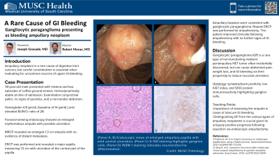
Introduction: Ampullary neoplasm is a rare cause of digestive tract cancers, but careful consideration is essential when evaluating for uncommon sources of upper GI bleeding. The most common types of ampullary neoplasm include adenoma, adenocarcinoma, and neuroendocrine tumor (NET). Gangliocytic paraganglioma (GP) is a rare type of non-functioning indolent periampullary NET tumor that are often incidentally discovered, but can also cause abdominal pain, weight loss, and gastrointestinal (GI) bleeding via their propensity to induce mucosal ulceration. Distinguishing GP from the various types of ampullary neoplasms is crucial given its uniquely positive prognosis following resection via endoscopic ampullectomy.
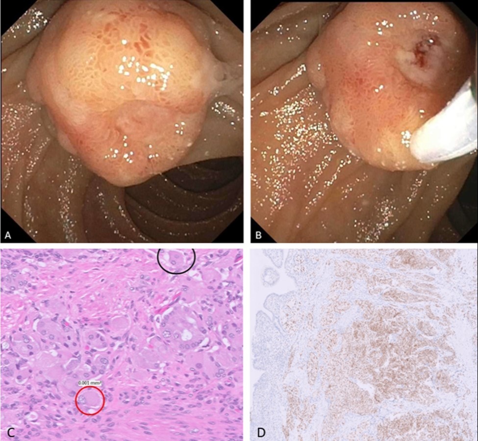
Case Description/Methods: A 78-year-old male with a history of chronic kidney disease presented with worsening dyspnea over a one-week period in the setting of melena and two episodes of coffee-ground emesis. The patient was hemodynamically stable at time of admission. Physical examination revealed a well-appearing elderly male with conjunctival pallor, no signs of jaundice, and a soft, non-tender abdominal exam. Exam was otherwise unremarkable. Initial labs revealed a hemoglobin level of 10 gm/dL from a prior baseline of 14 gm/dL, elevated BUN/Cr ratio of 28, and stable baseline creatinine 1.5 mg/dL. Forward-viewing endoscopy showed an enlarged erythematous ampulla with possible ulceration. MRCP revealed an enlarged 1.3 cm ampulla with no evidence of distant metastasis. ERCP was performed and revealed a major papilla measuring 1.5 cm with ulceration of the central part of the papilla. Multiple biopsies were obtained from the ampulla and were consistent with diagnosis of GP. Repeat ERCP was performed for ampullectomy. The patient improved clinically following ampullectomy with no further signs of GI bleeding. Subsequent surveillance endoscopy two months later showed no evidence of residual tumor.
Discussion: Gangliocytic paraganglioma (GP) is a rare tumor that accounts for a small portion of duodenal NETs and may cause significant GI bleeding. It can be distinguished by its characteristic histologic features, including synaptophysin positivity, low Ki67 index, and S100 protein immunoreactivity highlighting ganglion cells. This case highlights the efficacy of endoscopic ampullectomy in the management of GP. It also serves as a lesson to endoscopists in training to illustrate the importance of assessing the ampulla in cases of obscure GI bleeding.

Disclosures:
Joseph Granade, MD, Robert A. Moran, MD. P0812 - Gangliocytic Paraganglioma: Ampullary Neoplasm as a Rare Cause of Upper Gastrointestinal Bleeding, ACG 2024 Annual Scientific Meeting Abstracts. Philadelphia, PA: American College of Gastroenterology.
Medical University of South Carolina, Charleston, SC
Introduction: Ampullary neoplasm is a rare cause of digestive tract cancers, but careful consideration is essential when evaluating for uncommon sources of upper GI bleeding. The most common types of ampullary neoplasm include adenoma, adenocarcinoma, and neuroendocrine tumor (NET). Gangliocytic paraganglioma (GP) is a rare type of non-functioning indolent periampullary NET tumor that are often incidentally discovered, but can also cause abdominal pain, weight loss, and gastrointestinal (GI) bleeding via their propensity to induce mucosal ulceration. Distinguishing GP from the various types of ampullary neoplasms is crucial given its uniquely positive prognosis following resection via endoscopic ampullectomy.
Case Description/Methods: A 78-year-old male with a history of chronic kidney disease presented with worsening dyspnea over a one-week period in the setting of melena and two episodes of coffee-ground emesis. The patient was hemodynamically stable at time of admission. Physical examination revealed a well-appearing elderly male with conjunctival pallor, no signs of jaundice, and a soft, non-tender abdominal exam. Exam was otherwise unremarkable. Initial labs revealed a hemoglobin level of 10 gm/dL from a prior baseline of 14 gm/dL, elevated BUN/Cr ratio of 28, and stable baseline creatinine 1.5 mg/dL. Forward-viewing endoscopy showed an enlarged erythematous ampulla with possible ulceration. MRCP revealed an enlarged 1.3 cm ampulla with no evidence of distant metastasis. ERCP was performed and revealed a major papilla measuring 1.5 cm with ulceration of the central part of the papilla. Multiple biopsies were obtained from the ampulla and were consistent with diagnosis of GP. Repeat ERCP was performed for ampullectomy. The patient improved clinically following ampullectomy with no further signs of GI bleeding. Subsequent surveillance endoscopy two months later showed no evidence of residual tumor.
Discussion: Gangliocytic paraganglioma (GP) is a rare tumor that accounts for a small portion of duodenal NETs and may cause significant GI bleeding. It can be distinguished by its characteristic histologic features, including synaptophysin positivity, low Ki67 index, and S100 protein immunoreactivity highlighting ganglion cells. This case highlights the efficacy of endoscopic ampullectomy in the management of GP. It also serves as a lesson to endoscopists in training to illustrate the importance of assessing the ampulla in cases of obscure GI bleeding.

Figure: (Panel A, B) Endoscopic views of enlarged ampullary papilla with mild central ulceration. (Panel C) S-100 staining highlights ganglion cells. (Panel D) INSM-1 staining indicates neuroendocrine differentiation.
Disclosures:
Joseph Granade indicated no relevant financial relationships.
Robert Moran: Cook Medical – Consultant.
Joseph Granade, MD, Robert A. Moran, MD. P0812 - Gangliocytic Paraganglioma: Ampullary Neoplasm as a Rare Cause of Upper Gastrointestinal Bleeding, ACG 2024 Annual Scientific Meeting Abstracts. Philadelphia, PA: American College of Gastroenterology.

