Sunday Poster Session
Category: Interventional Endoscopy
P1056 - Efficacy of EUS With Miniprobe in Evaluating Right-Sided Colonic Lesions
Sunday, October 27, 2024
3:30 PM - 7:00 PM ET
Location: Exhibit Hall E

Has Audio
- DK
Daniel Kurtz, DO
Mount Sinai South Nassau, Icahn School of Medicine at Mount Sinai
Oceanside, NY
Presenting Author(s)
Neal Shah, DO1, Mahnoor Khan, DO1, Daniel Kurtz, DO1, Samuel Huang, MD1, Pranay Srivastava, MD1, Frank Gress, MD2
1Mount Sinai South Nassau, Icahn School of Medicine at Mount Sinai, Oceanside, NY; 2Mount Sinai South Nassau, Icahn School of Medicine at Mount Sinai, Bellmore, NY
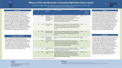
Introduction: Traditionally, colonic lesions found from the cecum through the hepatic flexure required surgical colon resection. With the development of advanced endoscopic techniques, such as endoscopic mucosal resection (EMR), there may be a less invasive way to treat right sided colonic lesions up to a certain size, avoiding the necessity of surgery. Endoscopic ultrasound (EUS) allows for high resolution imaging with the ability to assess tumor depth. Currently, multi-detector computed tomography (MDCT) is the main imaging modality for staging, however its accuracy is limited for lower stage lesions and tumor depth. EUS is already used for evaluating and staging rectal lesions, however, there is no current guidance on staging right sided colonic lesions.
Methods: This is a retrospective study that examined seven patients who underwent EUS of right sided colonic lesions between 2018 through present date by using miniprobe ultrasound. Originally, there was a pool of 41 patients who underwent lower EUS and was narrowed to include those who underwent EUS of right sided colonic lesions. This is a pilot study to investigate the efficacy of miniprobe ultrasound to identify the depth of invasion and diagnosis of right sided lesions.
Results: From among the cohort of 7 individuals, 2 lesions were found in the cecum, 2 in the ascending colon, 2 in the ileocecal valve, and 1 in the hepatic flexure. 100% of the 7 individuals that had an endoscopic ultrasound with miniprobe were useful in diagnosing lesions that found 4 lipomas, 2 adenomas, and 1 normal tissue. These lesions ranged in a variety of sizes from 3mm to 8cm. Because of the findings from the miniprobe, 5 individuals required no further intervention, 1 required robotic ileocolic resection, and 1 required follow up EMR.
Discussion: Our research showed that in all cases the EUS with use of miniprobe was successful in delineating the borders of the lesion. Additionally, in one case, EMR was completed after delineating borders with EUS and allowed for the complete resection of the lesion with clean margins. Although the sample size was small, we conclude that EUS may be an effective method for evaluation of depth of invasion and diagnosis of right sided lesions. Future studies will continue to examine patients with right sided lesions, especially those that are cancerous/high grade dysplasia to evaluate whether EUS can successfully be used to delineate those lesions and guide effective treatment with EMR with a number needed to treat of 10.
Note: The table for this abstract can be viewed in the ePoster Gallery section of the ACG 2024 ePoster Site or in The American Journal of Gastroenterology's abstract supplement issue, both of which will be available starting October 27, 2024.
Disclosures:
Neal Shah, DO1, Mahnoor Khan, DO1, Daniel Kurtz, DO1, Samuel Huang, MD1, Pranay Srivastava, MD1, Frank Gress, MD2. P1056 - Efficacy of EUS With Miniprobe in Evaluating Right-Sided Colonic Lesions, ACG 2024 Annual Scientific Meeting Abstracts. Philadelphia, PA: American College of Gastroenterology.
1Mount Sinai South Nassau, Icahn School of Medicine at Mount Sinai, Oceanside, NY; 2Mount Sinai South Nassau, Icahn School of Medicine at Mount Sinai, Bellmore, NY
Introduction: Traditionally, colonic lesions found from the cecum through the hepatic flexure required surgical colon resection. With the development of advanced endoscopic techniques, such as endoscopic mucosal resection (EMR), there may be a less invasive way to treat right sided colonic lesions up to a certain size, avoiding the necessity of surgery. Endoscopic ultrasound (EUS) allows for high resolution imaging with the ability to assess tumor depth. Currently, multi-detector computed tomography (MDCT) is the main imaging modality for staging, however its accuracy is limited for lower stage lesions and tumor depth. EUS is already used for evaluating and staging rectal lesions, however, there is no current guidance on staging right sided colonic lesions.
Methods: This is a retrospective study that examined seven patients who underwent EUS of right sided colonic lesions between 2018 through present date by using miniprobe ultrasound. Originally, there was a pool of 41 patients who underwent lower EUS and was narrowed to include those who underwent EUS of right sided colonic lesions. This is a pilot study to investigate the efficacy of miniprobe ultrasound to identify the depth of invasion and diagnosis of right sided lesions.
Results: From among the cohort of 7 individuals, 2 lesions were found in the cecum, 2 in the ascending colon, 2 in the ileocecal valve, and 1 in the hepatic flexure. 100% of the 7 individuals that had an endoscopic ultrasound with miniprobe were useful in diagnosing lesions that found 4 lipomas, 2 adenomas, and 1 normal tissue. These lesions ranged in a variety of sizes from 3mm to 8cm. Because of the findings from the miniprobe, 5 individuals required no further intervention, 1 required robotic ileocolic resection, and 1 required follow up EMR.
Discussion: Our research showed that in all cases the EUS with use of miniprobe was successful in delineating the borders of the lesion. Additionally, in one case, EMR was completed after delineating borders with EUS and allowed for the complete resection of the lesion with clean margins. Although the sample size was small, we conclude that EUS may be an effective method for evaluation of depth of invasion and diagnosis of right sided lesions. Future studies will continue to examine patients with right sided lesions, especially those that are cancerous/high grade dysplasia to evaluate whether EUS can successfully be used to delineate those lesions and guide effective treatment with EMR with a number needed to treat of 10.
Note: The table for this abstract can be viewed in the ePoster Gallery section of the ACG 2024 ePoster Site or in The American Journal of Gastroenterology's abstract supplement issue, both of which will be available starting October 27, 2024.
Disclosures:
Neal Shah indicated no relevant financial relationships.
Mahnoor Khan indicated no relevant financial relationships.
Daniel Kurtz indicated no relevant financial relationships.
Samuel Huang indicated no relevant financial relationships.
Pranay Srivastava indicated no relevant financial relationships.
Frank Gress indicated no relevant financial relationships.
Neal Shah, DO1, Mahnoor Khan, DO1, Daniel Kurtz, DO1, Samuel Huang, MD1, Pranay Srivastava, MD1, Frank Gress, MD2. P1056 - Efficacy of EUS With Miniprobe in Evaluating Right-Sided Colonic Lesions, ACG 2024 Annual Scientific Meeting Abstracts. Philadelphia, PA: American College of Gastroenterology.
