Sunday Poster Session
Category: Interventional Endoscopy
P1102 - A Rare Roadblock: A Case of Sporadic Ampullary Tubulovillous Adenoma Leading to Acute Pancreatitis
Sunday, October 27, 2024
3:30 PM - 7:00 PM ET
Location: Exhibit Hall E

Has Audio

Anvit D. Reddy, DO
University of Florida College of Medicine
Jacksonville, FL
Presenting Author(s)
Anvit D. Reddy, DO1, Falguni Patel, DO1, Niyati Patel, DO1, Nadim A. Qadir, DO2, Bruno Ribeiro, MD1
1University of Florida College of Medicine, Jacksonville, FL; 2University of Florida College of Medicine, Windermere, FL
Introduction: Duodenal adenomas (DA) are commonly found in familial conditions such as Gardner syndrome or Familial Adenomatous Polyposis. In the absence of underlying conditions, these adenomas are classified as sporadic duodenal adenomas (SDA) which are uncommon. We present a case of an SDA leading to acute pancreatitis.
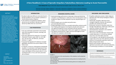
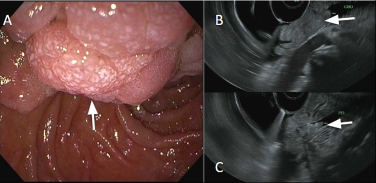
Case Description/Methods: A 67-year-old female presented to the hospital with chief complaints of nausea, vomiting, and abdominal pain. Labs were significant for potassium 2.9 mmol/L, chloride 87 mmol/L, lipase 1067 U/L, AST 588 IU/L, ALT 222 IU/L, ALP 296 IU/L, and total bilirubin 2.1 mg/dL. Computed Tomography (CT) abdomen revealed gallbladder distention and severe dilation of the biliary ducts. A magnetic resonance cholangiopancreatography showed a 2.5 centimeter (cm) hypointense mass at the ampulla that projected into the duodenal lumen and dilation of the common bile (1.1 cm), intrahepatic bile (1.4 cm), and main pancreatic (0.5 cm) ducts. Gastroenterology performed an endoscopic ultrasound (EUS) that revealed a non-bleeding 6-cm adenoma in the second portion of the duodenum (Figure 1A) with projection into the ampulla (Figure 1B and C). General Surgery was consulted and performed a pancreaticoduodenectomy. Surgical pathology of the mass revealed tubulovillous adenoma with foci of high-grade dysplasia involving the ampulla with extension into adjacent duodenal mucosa and distal common bile duct. Margins and surrounding lymph nodes were negative for metastasis. The patient recovered from surgery and was discharged to home with plans for outpatient colonoscopy.
Discussion: Ampullary adenomas possess a high malignant potential with 79% having histological evidence of carcinoma. Historically, ampullary adenomas were managed surgically, however, the operative mortality was noted to be 1%-9%. Endoscopic removal of ampullary adenomas through ampullectomy, endoscopic submucosal dissection, ablative therapies, and sphincterotomies represent a non-standard approach to management. The success rates of endoscopic removal with smaller adenomas (< 2 cm) and non-dilated ducts have more favorable outcomes. In this patient, it was decided to proceed with surgery due to the size of the adenoma and the presence of intraductal projections within the biliary and pancreatic ducts. Patients with duodenal adenomas have a significant association with colorectal adenomas, so colonoscopy surveillance is recommended.

Disclosures:
Anvit D. Reddy, DO1, Falguni Patel, DO1, Niyati Patel, DO1, Nadim A. Qadir, DO2, Bruno Ribeiro, MD1. P1102 - A Rare Roadblock: A Case of Sporadic Ampullary Tubulovillous Adenoma Leading to Acute Pancreatitis, ACG 2024 Annual Scientific Meeting Abstracts. Philadelphia, PA: American College of Gastroenterology.
1University of Florida College of Medicine, Jacksonville, FL; 2University of Florida College of Medicine, Windermere, FL
Introduction: Duodenal adenomas (DA) are commonly found in familial conditions such as Gardner syndrome or Familial Adenomatous Polyposis. In the absence of underlying conditions, these adenomas are classified as sporadic duodenal adenomas (SDA) which are uncommon. We present a case of an SDA leading to acute pancreatitis.
Case Description/Methods: A 67-year-old female presented to the hospital with chief complaints of nausea, vomiting, and abdominal pain. Labs were significant for potassium 2.9 mmol/L, chloride 87 mmol/L, lipase 1067 U/L, AST 588 IU/L, ALT 222 IU/L, ALP 296 IU/L, and total bilirubin 2.1 mg/dL. Computed Tomography (CT) abdomen revealed gallbladder distention and severe dilation of the biliary ducts. A magnetic resonance cholangiopancreatography showed a 2.5 centimeter (cm) hypointense mass at the ampulla that projected into the duodenal lumen and dilation of the common bile (1.1 cm), intrahepatic bile (1.4 cm), and main pancreatic (0.5 cm) ducts. Gastroenterology performed an endoscopic ultrasound (EUS) that revealed a non-bleeding 6-cm adenoma in the second portion of the duodenum (Figure 1A) with projection into the ampulla (Figure 1B and C). General Surgery was consulted and performed a pancreaticoduodenectomy. Surgical pathology of the mass revealed tubulovillous adenoma with foci of high-grade dysplasia involving the ampulla with extension into adjacent duodenal mucosa and distal common bile duct. Margins and surrounding lymph nodes were negative for metastasis. The patient recovered from surgery and was discharged to home with plans for outpatient colonoscopy.
Discussion: Ampullary adenomas possess a high malignant potential with 79% having histological evidence of carcinoma. Historically, ampullary adenomas were managed surgically, however, the operative mortality was noted to be 1%-9%. Endoscopic removal of ampullary adenomas through ampullectomy, endoscopic submucosal dissection, ablative therapies, and sphincterotomies represent a non-standard approach to management. The success rates of endoscopic removal with smaller adenomas (< 2 cm) and non-dilated ducts have more favorable outcomes. In this patient, it was decided to proceed with surgery due to the size of the adenoma and the presence of intraductal projections within the biliary and pancreatic ducts. Patients with duodenal adenomas have a significant association with colorectal adenomas, so colonoscopy surveillance is recommended.

Figure: (A): 6-cm adenoma in the second portion of the duodenum with projection into the ampulla
(B): Endoscopic ultrasound (EUS) demonstrating adenoma invading into the common bile duct
(C): Endoscopic ultrasound (EUS) demonstrating adenoma invading into the pancreatic duct
(B): Endoscopic ultrasound (EUS) demonstrating adenoma invading into the common bile duct
(C): Endoscopic ultrasound (EUS) demonstrating adenoma invading into the pancreatic duct
Disclosures:
Anvit Reddy indicated no relevant financial relationships.
Falguni Patel indicated no relevant financial relationships.
Niyati Patel indicated no relevant financial relationships.
Nadim Qadir indicated no relevant financial relationships.
Bruno Ribeiro indicated no relevant financial relationships.
Anvit D. Reddy, DO1, Falguni Patel, DO1, Niyati Patel, DO1, Nadim A. Qadir, DO2, Bruno Ribeiro, MD1. P1102 - A Rare Roadblock: A Case of Sporadic Ampullary Tubulovillous Adenoma Leading to Acute Pancreatitis, ACG 2024 Annual Scientific Meeting Abstracts. Philadelphia, PA: American College of Gastroenterology.
