Sunday Poster Session
Category: Interventional Endoscopy
P1107 - Complexity of Inflammatory Fibroid Polyps
Sunday, October 27, 2024
3:30 PM - 7:00 PM ET
Location: Exhibit Hall E

Has Audio
- SE
Sunanda Ellina, MD
Creighton University School of Medicine
Phoenix, AZ
Presenting Author(s)
Sunanda Ellina, MD1, Aida Rezaie, MD2, Savio Reddymasu, MBBS, FACG3
1Creighton University School of Medicine, Phoenix, AZ; 2Creighton University, Phoenix, AZ; 3Creighton University Medical Center, Phoenix, AZ
Introduction: Inflammatory fibroid polyps are rare, idiopathic and benign lesions that can be found throughout the gastrointestinal tract. They are most commonly located in the stomach and the small intestine. Histologically, they are submucosal in origin and were previously considered reactive in nature. From an immunohistochemical standpoint, IFPs are typically positive for CD34, smooth muscle actin and CD68 and negative for CD117, S100 protein and cytokeratin and should be differentiated from other spindle cell lesions like GISTs, schwannomas and inflammatory myofibroblastic tumors via this staining method. Depending on the size of the polyp, significant bleeding, anemia and small bowel obstruction due to intussusception may occur. Therefore, surgical or endoscopic treatment may be recommended in symptomatic patients. We present a case of an inflammatory fibroid polyp detected on capsule endoscopy, confirmed and endoscopically removed by balloon enteroscopy.
Case Description/Methods: 81 year old man with a history of Barrett’s esophagus and pancreatic insufficiency presented for evaluation of iron deficiency anemia and weight loss. He denied any active signs of bleeding. He had a negative EGD and Colonoscopy. Video capsule endoscopy (VCE) revealed a mass in the distal ileum. Retrograde balloon enteroscopy showed a 2.5 cm polypoid lesion with superficial ulceration 25 cm from the ileocecal valve. The polyp was removed en-bloc with hot snare cautery, and India Ink tattoo was used to demarcate the location. Pathology showed a focally ulcerated polypoid lesion with reactive atypia and focal surface ulceration. The underlying stroma appeared to be lymphoid-rich with interspersed eosinophils and thick-walled blood vessels. The polyp was CD34, smooth muscle actin, muscle specific actin, desmin and CD117 negative on immunohistochemical staining. Genomic testing was unrevealing for any mutations in PDGFRA, and most suggestive of an inflammatory fibroid polyp.
Discussion: The diagnosis and management of ileal polyps can be complex, and requires expert pathological interpretation from a GI pathologist to differentiate it from neoplastic lesions. Most IFP’s are diagnosed after surgical removal of the small bowel after intussusception or obstruction, and range from 2 cm to 5 cm in diameter. We highlight a unique case of IFP diagnosed by VCE and feasibility of deep enteroscopy as a minimally invasive alternative to surgical resection to safely remove these lesions (including larger lesions).

Disclosures:
Sunanda Ellina, MD1, Aida Rezaie, MD2, Savio Reddymasu, MBBS, FACG3. P1107 - Complexity of Inflammatory Fibroid Polyps, ACG 2024 Annual Scientific Meeting Abstracts. Philadelphia, PA: American College of Gastroenterology.
1Creighton University School of Medicine, Phoenix, AZ; 2Creighton University, Phoenix, AZ; 3Creighton University Medical Center, Phoenix, AZ
Introduction: Inflammatory fibroid polyps are rare, idiopathic and benign lesions that can be found throughout the gastrointestinal tract. They are most commonly located in the stomach and the small intestine. Histologically, they are submucosal in origin and were previously considered reactive in nature. From an immunohistochemical standpoint, IFPs are typically positive for CD34, smooth muscle actin and CD68 and negative for CD117, S100 protein and cytokeratin and should be differentiated from other spindle cell lesions like GISTs, schwannomas and inflammatory myofibroblastic tumors via this staining method. Depending on the size of the polyp, significant bleeding, anemia and small bowel obstruction due to intussusception may occur. Therefore, surgical or endoscopic treatment may be recommended in symptomatic patients. We present a case of an inflammatory fibroid polyp detected on capsule endoscopy, confirmed and endoscopically removed by balloon enteroscopy.
Case Description/Methods: 81 year old man with a history of Barrett’s esophagus and pancreatic insufficiency presented for evaluation of iron deficiency anemia and weight loss. He denied any active signs of bleeding. He had a negative EGD and Colonoscopy. Video capsule endoscopy (VCE) revealed a mass in the distal ileum. Retrograde balloon enteroscopy showed a 2.5 cm polypoid lesion with superficial ulceration 25 cm from the ileocecal valve. The polyp was removed en-bloc with hot snare cautery, and India Ink tattoo was used to demarcate the location. Pathology showed a focally ulcerated polypoid lesion with reactive atypia and focal surface ulceration. The underlying stroma appeared to be lymphoid-rich with interspersed eosinophils and thick-walled blood vessels. The polyp was CD34, smooth muscle actin, muscle specific actin, desmin and CD117 negative on immunohistochemical staining. Genomic testing was unrevealing for any mutations in PDGFRA, and most suggestive of an inflammatory fibroid polyp.
Discussion: The diagnosis and management of ileal polyps can be complex, and requires expert pathological interpretation from a GI pathologist to differentiate it from neoplastic lesions. Most IFP’s are diagnosed after surgical removal of the small bowel after intussusception or obstruction, and range from 2 cm to 5 cm in diameter. We highlight a unique case of IFP diagnosed by VCE and feasibility of deep enteroscopy as a minimally invasive alternative to surgical resection to safely remove these lesions (including larger lesions).

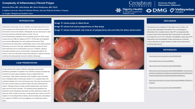
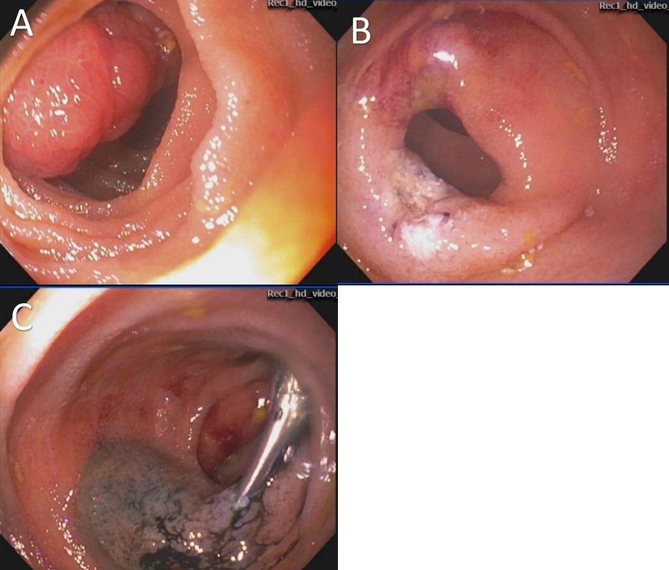
Figure: Image 'A' shows polyp in distal ileum
Image 'B' shows hot snare polypectomy of ileal polyp
Image 'C' shows hemostatic clip closure of polypectomy site and India Ink tattoo demarcation
Image 'B' shows hot snare polypectomy of ileal polyp
Image 'C' shows hemostatic clip closure of polypectomy site and India Ink tattoo demarcation
Disclosures:
Sunanda Ellina indicated no relevant financial relationships.
Aida Rezaie indicated no relevant financial relationships.
Savio Reddymasu indicated no relevant financial relationships.
Sunanda Ellina, MD1, Aida Rezaie, MD2, Savio Reddymasu, MBBS, FACG3. P1107 - Complexity of Inflammatory Fibroid Polyps, ACG 2024 Annual Scientific Meeting Abstracts. Philadelphia, PA: American College of Gastroenterology.
