Sunday Poster Session
Category: Interventional Endoscopy
P1116 - Hemostasis of Malignant Gastrointestinal Lesions With Over-The-Scope Clips: A Case Series
Sunday, October 27, 2024
3:30 PM - 7:00 PM ET
Location: Exhibit Hall E

Has Audio

Sriya Bhumi, MD, MBA
New York Presbyterian Queens
Flushing, NY
Presenting Author(s)
Award: Presidential Poster Award
Constantine Fisher, MD1, Sriya Bhumi, MD, MBA2, Syed Hussain, MD1, Sang Hoon Kim, MD1
1New York-Presbyterian/Queens, Flushing, NY; 2New York Presbyterian Queens, Flushing, NY
Introduction: Successful hemostasis of gastrointestinal masses depends on factors including lesion size, ulcer severity, and endoscopist skill level. Conventional techniques for hemostasis include vasoconstrictor administration, cauterization, and through-the-scope clipping.
In cases where conventional hemostatic methods fail, over-the-scope clips (OTSCs) may be used. OTSCs were initially indicated for closure of gastrotomies and natural orifice surgery procedures. There is a paucity of literature demonstrating use of OTSC for hemostasis, especially with regard to bleeding malignant lesions. The aim of this case series is to illustrate successful hemostasis of malignant lesions with the use OTSCs.
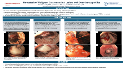
Case Description/Methods: A 73-year-old female presented with hematemesis. Hemoglobin was 6.9 g/dL. An endoscopic gastroduodenoscopy (EGD) revealed a 5cm ulcerated, submucosal gastric mass with active bleeding (Fig. 1a). The area was injected with epinephrine and one OTSC was deployed with hemostasis (Fig. 1b). Biopsies were positive for gastrointestinal stromal tumor, for which the patient underwent gastric wedge resection.
A 59-year-old man presented with melena. EGD showed an actively bleeding duodenal lesion. After injection with epinephrine, a visible vessel within an ulcer was revealed, atop a 3cm submucosal mass (Fig. 2a). One OTSC achieved hemostasis (Fig. 2b). Endoscopic ultrasound with biopsies revealed high grade B-cell lymphoma.
A 51-year-old male presented with a hemoglobin of 5.8 g/dL. EGD showed a 4cm gastric ulcer with spurting hemorrhage on the lesser curvature of the stomach (Fig. 3a). An OTSC was placed with hemostasis (Fig. 3b). Biopsies were positive for moderately differentiated adenocarcinoma, requiring subtotal gastrectomy.
A 61-year-old male presented with hematochezia. Hemoglobin was 7.4 g/dL. Colonoscopy revealed a 4cm bleeding cecal mass with biopsy proven adenocarcinoma (Fig. 4a). The area was injected with epinephrine and an OTSC was used (Fig. 4b). The patient had no further bleeding and underwent a right hemicolectomy after waiting for antiplatelet agent effect to dissipate.
Discussion: We describe successful hemostasis involving a variety of bleeding malignant lesions with OTSCs. Given lesion characteristics (size, placement, etc), conventional means of hemostasis would likely not be successful. Utilization of an available tool in a novel fashion provides additional means of endoscopic therapy, allowing for stabilization of patients with the ability to plan subsequent management.

Disclosures:
Constantine Fisher, MD1, Sriya Bhumi, MD, MBA2, Syed Hussain, MD1, Sang Hoon Kim, MD1. P1116 - Hemostasis of Malignant Gastrointestinal Lesions With Over-The-Scope Clips: A Case Series, ACG 2024 Annual Scientific Meeting Abstracts. Philadelphia, PA: American College of Gastroenterology.
Constantine Fisher, MD1, Sriya Bhumi, MD, MBA2, Syed Hussain, MD1, Sang Hoon Kim, MD1
1New York-Presbyterian/Queens, Flushing, NY; 2New York Presbyterian Queens, Flushing, NY
Introduction: Successful hemostasis of gastrointestinal masses depends on factors including lesion size, ulcer severity, and endoscopist skill level. Conventional techniques for hemostasis include vasoconstrictor administration, cauterization, and through-the-scope clipping.
In cases where conventional hemostatic methods fail, over-the-scope clips (OTSCs) may be used. OTSCs were initially indicated for closure of gastrotomies and natural orifice surgery procedures. There is a paucity of literature demonstrating use of OTSC for hemostasis, especially with regard to bleeding malignant lesions. The aim of this case series is to illustrate successful hemostasis of malignant lesions with the use OTSCs.
Case Description/Methods: A 73-year-old female presented with hematemesis. Hemoglobin was 6.9 g/dL. An endoscopic gastroduodenoscopy (EGD) revealed a 5cm ulcerated, submucosal gastric mass with active bleeding (Fig. 1a). The area was injected with epinephrine and one OTSC was deployed with hemostasis (Fig. 1b). Biopsies were positive for gastrointestinal stromal tumor, for which the patient underwent gastric wedge resection.
A 59-year-old man presented with melena. EGD showed an actively bleeding duodenal lesion. After injection with epinephrine, a visible vessel within an ulcer was revealed, atop a 3cm submucosal mass (Fig. 2a). One OTSC achieved hemostasis (Fig. 2b). Endoscopic ultrasound with biopsies revealed high grade B-cell lymphoma.
A 51-year-old male presented with a hemoglobin of 5.8 g/dL. EGD showed a 4cm gastric ulcer with spurting hemorrhage on the lesser curvature of the stomach (Fig. 3a). An OTSC was placed with hemostasis (Fig. 3b). Biopsies were positive for moderately differentiated adenocarcinoma, requiring subtotal gastrectomy.
A 61-year-old male presented with hematochezia. Hemoglobin was 7.4 g/dL. Colonoscopy revealed a 4cm bleeding cecal mass with biopsy proven adenocarcinoma (Fig. 4a). The area was injected with epinephrine and an OTSC was used (Fig. 4b). The patient had no further bleeding and underwent a right hemicolectomy after waiting for antiplatelet agent effect to dissipate.
Discussion: We describe successful hemostasis involving a variety of bleeding malignant lesions with OTSCs. Given lesion characteristics (size, placement, etc), conventional means of hemostasis would likely not be successful. Utilization of an available tool in a novel fashion provides additional means of endoscopic therapy, allowing for stabilization of patients with the ability to plan subsequent management.

Figure: Figure 1a. A 5cm ulcerated, submucosal mass with active bleeding.
Figure 1b. One OTSC was deployed on the submucosal mass with successful hemostasis.
Figure 2a. A visible vessel within an ulcer, atop a 3cm submucosal mass
Figure 2b. One OTSC deployed over submucosal lesion with hemostasis.
Figure 3a. A 4cm gastric ulcer with spurting hemorrhage on the lesser curvature of the stomach.
Figure 3b. An OTSC placed over gastric ulcer with cessation of spurting bleed.
Figure 4a. A 4cm bleeding cecal mass with biopsy proven adenocarcinoma.
Figure 4b. An OTSC deployed on bleeding cecal mass with hemostasis.
Disclosures:
Constantine Fisher indicated no relevant financial relationships.
Sriya Bhumi indicated no relevant financial relationships.
Syed Hussain indicated no relevant financial relationships.
Sang Hoon Kim indicated no relevant financial relationships.
Constantine Fisher, MD1, Sriya Bhumi, MD, MBA2, Syed Hussain, MD1, Sang Hoon Kim, MD1. P1116 - Hemostasis of Malignant Gastrointestinal Lesions With Over-The-Scope Clips: A Case Series, ACG 2024 Annual Scientific Meeting Abstracts. Philadelphia, PA: American College of Gastroenterology.

