Sunday Poster Session
Category: Liver
P1308 - Metastatic Gastrointestinal Stromal Tumor Presenting as a Massive Multiloculated Liver Cyst: An Uncommon and Potentially Life-Threatening Condition
Sunday, October 27, 2024
3:30 PM - 7:00 PM ET
Location: Exhibit Hall E

Has Audio

Anjo Chacko, MD
Broward Health North
Deerfield Beach, FL
Presenting Author(s)
Laura Miranda, MD1, Anjo Chacko, MD2, Keshavi Mahesh, MD1, Mina Ayad, MD3, Satya Singh, MD4
1Broward Health North, Pompano Beach, FL; 2Broward Health North, Deerfield Beach, FL; 3Broward Health North, Fort Lauderdale, FL; 4Broward Health Medical Center, Fort Lauderdale, FL
Introduction: Gastrointestinal stromal tumors (GISTs) are the most prevalent type of mesenchymal tumors, originating from the interstitial cells of Cajal within the gastrointestinal tract. Despite advances in treatment, a significant number of patients with GIST will experience recurrence or metastasis, predominantly in the liver or peritoneum. Typically, GISTs manifest as solid masses. However, GISTs presenting with cystic morphology, especially within the liver, are exceedingly rare. Hereby, we describe an unusual case of GIST presenting as a massive complex septated liver cyst.
Case Description/Methods: A 55-year-old female with a history of GIST s/p partial gastrectomy and colectomy in 2020 presents with right upper quadrant abdominal pain radiating to the back, poor oral intake, nausea, and vomiting. Physical exam revealed right upper quadrant distention and firmness. Laboratory workup was notable for total bilirubin 3.5 mg/dL, ALT 47 U/L, and lipase 121 U/L. CT liver triple-phase scan demonstrated a multiloculated cystic lesion measuring 16.8 x 26.8 x 20.6 cm and small volume perihepatic ascites. This lesion occupied the entire left lobe and significant portion of the right lobe, extending to the left upper quadrant, causing mass effect and posterior displacement of the stomach, spleen, and mesenteric structures. Patient underwent a left hepatectomy with resection of a large abdominal cyst measuring over 20 cm. Intraoperative findings included a large cystic mass replacing the entire left lobe of the liver, attached to the diaphragm, stomach, and spleen. Histopathological analysis revealed a malignant neoplasm positive for calponin, CD117 (c-KIT), and DOG1, most compatible with recurrent GIST.
Discussion: This case illustrates an unusual presentation of recurrent GIST as a cystic liver lesion prior to any treatment. Typically, liver metastases from GIST are hypervascular and only become cystic following treatment with imatinib. Purely cystic metastases of GIST are exceedingly rare, with only a handful of reported cases. Current GIST risk stratification criteria include size, mitotic activity, and location. However, these criteria do not account for gross morphology, which might be crucial in cases like ours. Therefore, it is essential to have a broad differential diagnosis for cystic liver lesions in patients with a history of GIST. Further research is needed to determine the appropriateness of existing risk stratification systems in this patient population and guide optimal treatment strategies.

Disclosures:
Laura Miranda, MD1, Anjo Chacko, MD2, Keshavi Mahesh, MD1, Mina Ayad, MD3, Satya Singh, MD4. P1308 - Metastatic Gastrointestinal Stromal Tumor Presenting as a Massive Multiloculated Liver Cyst: An Uncommon and Potentially Life-Threatening Condition, ACG 2024 Annual Scientific Meeting Abstracts. Philadelphia, PA: American College of Gastroenterology.
1Broward Health North, Pompano Beach, FL; 2Broward Health North, Deerfield Beach, FL; 3Broward Health North, Fort Lauderdale, FL; 4Broward Health Medical Center, Fort Lauderdale, FL
Introduction: Gastrointestinal stromal tumors (GISTs) are the most prevalent type of mesenchymal tumors, originating from the interstitial cells of Cajal within the gastrointestinal tract. Despite advances in treatment, a significant number of patients with GIST will experience recurrence or metastasis, predominantly in the liver or peritoneum. Typically, GISTs manifest as solid masses. However, GISTs presenting with cystic morphology, especially within the liver, are exceedingly rare. Hereby, we describe an unusual case of GIST presenting as a massive complex septated liver cyst.
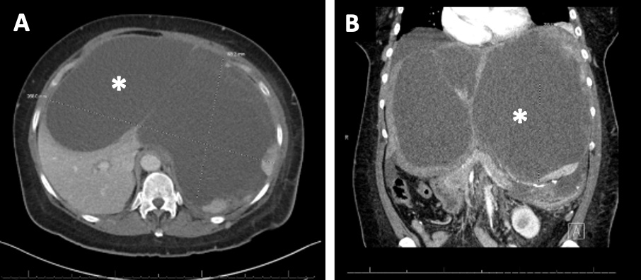
Case Description/Methods: A 55-year-old female with a history of GIST s/p partial gastrectomy and colectomy in 2020 presents with right upper quadrant abdominal pain radiating to the back, poor oral intake, nausea, and vomiting. Physical exam revealed right upper quadrant distention and firmness. Laboratory workup was notable for total bilirubin 3.5 mg/dL, ALT 47 U/L, and lipase 121 U/L. CT liver triple-phase scan demonstrated a multiloculated cystic lesion measuring 16.8 x 26.8 x 20.6 cm and small volume perihepatic ascites. This lesion occupied the entire left lobe and significant portion of the right lobe, extending to the left upper quadrant, causing mass effect and posterior displacement of the stomach, spleen, and mesenteric structures. Patient underwent a left hepatectomy with resection of a large abdominal cyst measuring over 20 cm. Intraoperative findings included a large cystic mass replacing the entire left lobe of the liver, attached to the diaphragm, stomach, and spleen. Histopathological analysis revealed a malignant neoplasm positive for calponin, CD117 (c-KIT), and DOG1, most compatible with recurrent GIST.
Discussion: This case illustrates an unusual presentation of recurrent GIST as a cystic liver lesion prior to any treatment. Typically, liver metastases from GIST are hypervascular and only become cystic following treatment with imatinib. Purely cystic metastases of GIST are exceedingly rare, with only a handful of reported cases. Current GIST risk stratification criteria include size, mitotic activity, and location. However, these criteria do not account for gross morphology, which might be crucial in cases like ours. Therefore, it is essential to have a broad differential diagnosis for cystic liver lesions in patients with a history of GIST. Further research is needed to determine the appropriateness of existing risk stratification systems in this patient population and guide optimal treatment strategies.

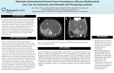
Figure: Axial (A) and coronal (B) CT liver triple phase images demonstrating a massive complex cystic mass throughout the liver.
Disclosures:
Laura Miranda indicated no relevant financial relationships.
Anjo Chacko indicated no relevant financial relationships.
Keshavi Mahesh indicated no relevant financial relationships.
Mina Ayad indicated no relevant financial relationships.
Satya Singh indicated no relevant financial relationships.
Laura Miranda, MD1, Anjo Chacko, MD2, Keshavi Mahesh, MD1, Mina Ayad, MD3, Satya Singh, MD4. P1308 - Metastatic Gastrointestinal Stromal Tumor Presenting as a Massive Multiloculated Liver Cyst: An Uncommon and Potentially Life-Threatening Condition, ACG 2024 Annual Scientific Meeting Abstracts. Philadelphia, PA: American College of Gastroenterology.
