Sunday Poster Session
Category: Liver
P1314 - Beaver Tail Liver: A Case Report on a Variant in Hepatic Morphology
Sunday, October 27, 2024
3:30 PM - 7:00 PM ET
Location: Exhibit Hall E

Has Audio
- IM
Ibrahim Mohammed, MD
Albany Medical Center
Albany, NY
Presenting Author(s)
Ibrahim Mohammed, MD, Ahmad Abulawi, MD, Khaled Elsokary, DO, Seth Richter, MD
Albany Medical Center, Albany, NY
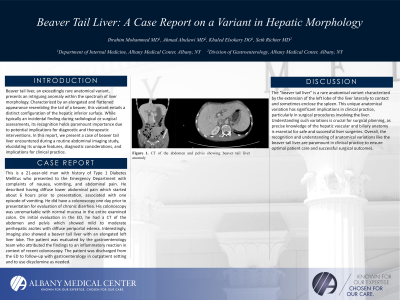
Introduction: Beaver tail liver, an exceedingly rare anatomical variant, presents an intriguing anomaly within the spectrum of liver morphology. Characterized by an elongated and flattened appearance resembling the tail of a beaver, this variant entails a distinct configuration of the hepatic inferior surface. While typically an incidental finding during radiological or surgical assessments, its recognition holds paramount importance due to potential implications for diagnostic and therapeutic interventions. In this report, we present a case of beaver tail liver encountered during a routine abdominal imaging study, elucidating its unique features, diagnostic considerations, and implications for clinical practice.
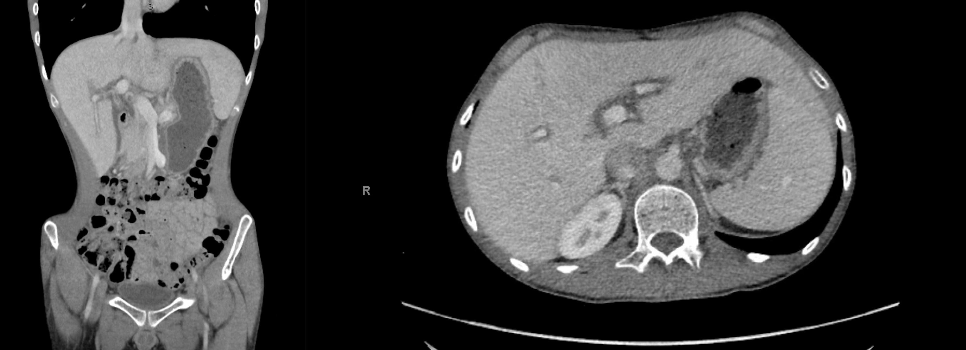
Case Description/Methods: This is a 21-year-old man with history of Type 1 Diabetes Mellitus who presented to the Emergency Department with complaints of nausea, vomiting, and abdominal pain. He described having diffuse lower abdominal pain which started about 6 hours prior to presentation, associated with one episode of vomiting. He did have a colonoscopy one day prior to presentation for evaluation of chronic diarrhea. His colonoscopy was unremarkable with normal mucosa in the entire examined colon. On initial evaluation in the ED, he had a CT of the abdomen and pelvis which showed mild to moderate perihepatic ascites with diffuse periportal edema. Interestingly, imaging also showed a beaver tail liver with an elongated left liver lobe. The patient was evaluated by the gastroenterology team who attributed the findings to an inflammatory reaction in context of recent colonoscopy. The patient was discharged from the ED to follow-up with gastroenterology in outpatient setting and to use dicyclomine as needed.
Discussion: The "beaver tail liver" is a rare anatomical variant characterized by the extension of the left lobe of the liver laterally to contact and sometimes enclose the spleen. This unique anatomical variation has significant implications in clinical practice, particularly in surgical procedures involving the liver. Understanding such variations is crucial for surgical planning, as precise knowledge of the hepatic vascular and biliary anatomy is essential for safe and successful liver surgeries. Overall, the recognition and understanding of anatomical variations like the beaver tail liver are paramount in clinical practice to ensure optimal patient care and successful surgical outcomes.

Disclosures:
Ibrahim Mohammed, MD, Ahmad Abulawi, MD, Khaled Elsokary, DO, Seth Richter, MD. P1314 - Beaver Tail Liver: A Case Report on a Variant in Hepatic Morphology, ACG 2024 Annual Scientific Meeting Abstracts. Philadelphia, PA: American College of Gastroenterology.
Albany Medical Center, Albany, NY
Introduction: Beaver tail liver, an exceedingly rare anatomical variant, presents an intriguing anomaly within the spectrum of liver morphology. Characterized by an elongated and flattened appearance resembling the tail of a beaver, this variant entails a distinct configuration of the hepatic inferior surface. While typically an incidental finding during radiological or surgical assessments, its recognition holds paramount importance due to potential implications for diagnostic and therapeutic interventions. In this report, we present a case of beaver tail liver encountered during a routine abdominal imaging study, elucidating its unique features, diagnostic considerations, and implications for clinical practice.
Case Description/Methods: This is a 21-year-old man with history of Type 1 Diabetes Mellitus who presented to the Emergency Department with complaints of nausea, vomiting, and abdominal pain. He described having diffuse lower abdominal pain which started about 6 hours prior to presentation, associated with one episode of vomiting. He did have a colonoscopy one day prior to presentation for evaluation of chronic diarrhea. His colonoscopy was unremarkable with normal mucosa in the entire examined colon. On initial evaluation in the ED, he had a CT of the abdomen and pelvis which showed mild to moderate perihepatic ascites with diffuse periportal edema. Interestingly, imaging also showed a beaver tail liver with an elongated left liver lobe. The patient was evaluated by the gastroenterology team who attributed the findings to an inflammatory reaction in context of recent colonoscopy. The patient was discharged from the ED to follow-up with gastroenterology in outpatient setting and to use dicyclomine as needed.
Discussion: The "beaver tail liver" is a rare anatomical variant characterized by the extension of the left lobe of the liver laterally to contact and sometimes enclose the spleen. This unique anatomical variation has significant implications in clinical practice, particularly in surgical procedures involving the liver. Understanding such variations is crucial for surgical planning, as precise knowledge of the hepatic vascular and biliary anatomy is essential for safe and successful liver surgeries. Overall, the recognition and understanding of anatomical variations like the beaver tail liver are paramount in clinical practice to ensure optimal patient care and successful surgical outcomes.

Figure: Figure 1. CT of the abdomen and pelvis showing beaver tail liver anomaly
Disclosures:
Ibrahim Mohammed indicated no relevant financial relationships.
Ahmad Abulawi indicated no relevant financial relationships.
Khaled Elsokary indicated no relevant financial relationships.
Seth Richter indicated no relevant financial relationships.
Ibrahim Mohammed, MD, Ahmad Abulawi, MD, Khaled Elsokary, DO, Seth Richter, MD. P1314 - Beaver Tail Liver: A Case Report on a Variant in Hepatic Morphology, ACG 2024 Annual Scientific Meeting Abstracts. Philadelphia, PA: American College of Gastroenterology.
