Sunday Poster Session
Category: Liver
P1333 - Diffuse Hepatic Hemangiomatosis in the Rear View
Sunday, October 27, 2024
3:30 PM - 7:00 PM ET
Location: Exhibit Hall E

Has Audio

Marilee Tiru-Vega, MD
VA Caribbean Healthcare System
San Juan, PR
Presenting Author(s)
Marilee Tirú-Vega, MD, Orlando Rodriguez-Amador, MD, Reyshley Ramos-Márquez, MD, Esteban Grovas-Cordovi, MD, Eduardo Acosta-Pumarejo, MD, Jose Martin-Ortiz, MD, FACG, Jaime Martinez-Souss, MD
VA Caribbean Healthcare System, San Juan, Puerto Rico
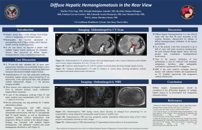
Introduction: Hepatic masses have a wide etiology from benign lesions to malignancy and metastatic disease. Radiography has become paramount in distinguishing most of these entities, and if obscure, further work-up should be pursued. In the case below, we describe a patient with multiple hepatic lesions worrisome for metastatic disease, later perceived as diffuse hepatic hemangiomatosis when examined retrospectively.
Case Description/Methods: A 79-year-old male admitted due to acute post-polypectomy bleeding was incidentally found to have mild elevation in transaminases warranting abdominal sonogram that showed multiple hepatic lesions. Abdominopelvic CT was then performed confirming hypodense, hepatic masses, largest measuring 14 cm transversely in the right hepatic lobe, with peripheral nodular enhancement. These masses were suspicious for hepatic metastasis from an unknown primary versus multicentric hepatocellular carcinoma. As part of the malignancy work-up, Chest CT and tumor markers were unremarkable (AFP 1.26, CA-19-9 18.3 and CEA 1.7). Recent colonoscopy was only pertinent for 3 tubular adenomatous polyps. Further characterization with MRI showed replacement of the hepatic parenchyma by innumerable round lesions, with low T1 and markedly high T2 signal intensity; as well as discontinuous peripheral nodular contrast enhancement with progressive filling of contrast in the delayed images. Imaging was consistent with multiple hepatic hemangiomas, with additional vertebral and costal hemangiomas identified as well.
Discussion: Diffuse hepatic hemangiomatosis is a rare clinical entity with less than 20 cases described in the existing literature, characterized by lobular or diffuse hemangiomatous infiltration of the hepatic parenchyma. As in this patient, it has been associated in up to half of cases with giant cavernous hemangiomas, the most common benign hepatic lesion, and may present with extrahepatic hemangiomatous involvement. Due to the massive infiltration of liver parenchyma, it may be confused with metastatic malignancy; therefore, adept radiographic examination is paramount for its diagnosis. MRI is the most sensitive technique showing lesions with indistinct borders and hyperintensity on T2 weighted sequencing with progressive contrast enhancement. In conclusion, diffuse hepatic hemangiomatosis should be considered in the differential diagnosis of multiple hepatic masses as its presentation may masquerade metastatic disease.

Disclosures:
Marilee Tirú-Vega, MD, Orlando Rodriguez-Amador, MD, Reyshley Ramos-Márquez, MD, Esteban Grovas-Cordovi, MD, Eduardo Acosta-Pumarejo, MD, Jose Martin-Ortiz, MD, FACG, Jaime Martinez-Souss, MD. P1333 - Diffuse Hepatic Hemangiomatosis in the Rear View, ACG 2024 Annual Scientific Meeting Abstracts. Philadelphia, PA: American College of Gastroenterology.
VA Caribbean Healthcare System, San Juan, Puerto Rico
Introduction: Hepatic masses have a wide etiology from benign lesions to malignancy and metastatic disease. Radiography has become paramount in distinguishing most of these entities, and if obscure, further work-up should be pursued. In the case below, we describe a patient with multiple hepatic lesions worrisome for metastatic disease, later perceived as diffuse hepatic hemangiomatosis when examined retrospectively.
Case Description/Methods: A 79-year-old male admitted due to acute post-polypectomy bleeding was incidentally found to have mild elevation in transaminases warranting abdominal sonogram that showed multiple hepatic lesions. Abdominopelvic CT was then performed confirming hypodense, hepatic masses, largest measuring 14 cm transversely in the right hepatic lobe, with peripheral nodular enhancement. These masses were suspicious for hepatic metastasis from an unknown primary versus multicentric hepatocellular carcinoma. As part of the malignancy work-up, Chest CT and tumor markers were unremarkable (AFP 1.26, CA-19-9 18.3 and CEA 1.7). Recent colonoscopy was only pertinent for 3 tubular adenomatous polyps. Further characterization with MRI showed replacement of the hepatic parenchyma by innumerable round lesions, with low T1 and markedly high T2 signal intensity; as well as discontinuous peripheral nodular contrast enhancement with progressive filling of contrast in the delayed images. Imaging was consistent with multiple hepatic hemangiomas, with additional vertebral and costal hemangiomas identified as well.
Discussion: Diffuse hepatic hemangiomatosis is a rare clinical entity with less than 20 cases described in the existing literature, characterized by lobular or diffuse hemangiomatous infiltration of the hepatic parenchyma. As in this patient, it has been associated in up to half of cases with giant cavernous hemangiomas, the most common benign hepatic lesion, and may present with extrahepatic hemangiomatous involvement. Due to the massive infiltration of liver parenchyma, it may be confused with metastatic malignancy; therefore, adept radiographic examination is paramount for its diagnosis. MRI is the most sensitive technique showing lesions with indistinct borders and hyperintensity on T2 weighted sequencing with progressive contrast enhancement. In conclusion, diffuse hepatic hemangiomatosis should be considered in the differential diagnosis of multiple hepatic masses as its presentation may masquerade metastatic disease.

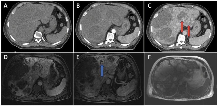
Figure: Figure 1:
1A: Abdominopelvic CT without contrast showing hepatomegaly with a coarse echotexture and multiple mass lesions, largest measuring 10.3 cm x 14 cm x 8.4 cm.
1B: Triphasic abdominopelvic CT with IV contrast in arterial phase showing multiple hepatic lesions.
1C: Triphasic abdominopelvic CT with IV contrast in venous phase showing peripheral, nodular, interrupted centripetal enhancement (red arrows).
1D: Abdominopelvic MRI during venous phase showing an enlarged liver (measuring 23 cm longitudinally), being replaced by innumerable round lesions of variable sizes.
1E: Abdominopelvic MRI showing peripheral nodular interrupted enhancement (blue arrow) which progress centripetally (inward) on delay images .
1F: Abdominopelvic MRI demonstrating T2 hyperintense lesions relative to liver parenchyma.
Disclosures:
Marilee Tirú-Vega indicated no relevant financial relationships.
Orlando Rodriguez-Amador indicated no relevant financial relationships.
Reyshley Ramos-Márquez indicated no relevant financial relationships.
Esteban Grovas-Cordovi indicated no relevant financial relationships.
Eduardo Acosta-Pumarejo indicated no relevant financial relationships.
Jose Martin-Ortiz indicated no relevant financial relationships.
Jaime Martinez-Souss indicated no relevant financial relationships.
Marilee Tirú-Vega, MD, Orlando Rodriguez-Amador, MD, Reyshley Ramos-Márquez, MD, Esteban Grovas-Cordovi, MD, Eduardo Acosta-Pumarejo, MD, Jose Martin-Ortiz, MD, FACG, Jaime Martinez-Souss, MD. P1333 - Diffuse Hepatic Hemangiomatosis in the Rear View, ACG 2024 Annual Scientific Meeting Abstracts. Philadelphia, PA: American College of Gastroenterology.
