Sunday Poster Session
Category: Liver
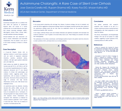
P1375 - Autoimmune Cholangitis: A Rare Case of Silent Liver Cirrhosis
Sunday, October 27, 2024
3:30 PM - 7:00 PM ET
Location: Exhibit Hall E

Has Audio
.jpg)
Jose Garcia-Corella, MD
Kern Medical Center
Bakersfield, CA
Presenting Author(s)
Jose Garcia-Corella, MD, Rupam Sharma, MD, Kasey Fox, DO, Ishaan Kalha, MD
Kern Medical Center, Bakersfield, CA
Introduction: Autoimmune cholangitis (AIC) is a variant syndrome of autoimmune hepatitis (AIH) which consists of a spectrum of diseases including small-duct primary sclerosing cholangitis (sdPSC), AMA-negative primary biliary cirrhosis (PBC), and IgG4 mediated sclerosing cholangitis.
The immune-mediated bile duct destruction can lead to chronic inflammation and irreversible complications. Here we present a rare case of an AIC variant resulting in cirrhosis in a female patient.
Case Description/Methods: A 41-year-old Hispanic female with no significant medical or social history, presented with one month of progressive abdominal distention, jaundice, peripheral edema, weakness, mild right upper quadrant abdominal pain, and weight loss. Initial workup was notable for abnormal liver function tests (ALT 67 U/L, AST 158 U/L, ALP 332 U/L) and direct hyperbilirubinemia (9.4 mg/dL). Abdominal ultrasound and CT scan showed evidence of cirrhosis with splenomegaly and collateral vessels, suggesting portal venous hypertension with no masses or other signs of obstruction. Noninvasive fibrosis testing showed stage F4 (cirrhosis) with high necrosis inflammatory activity score. EGD showed trace esophageal varices.
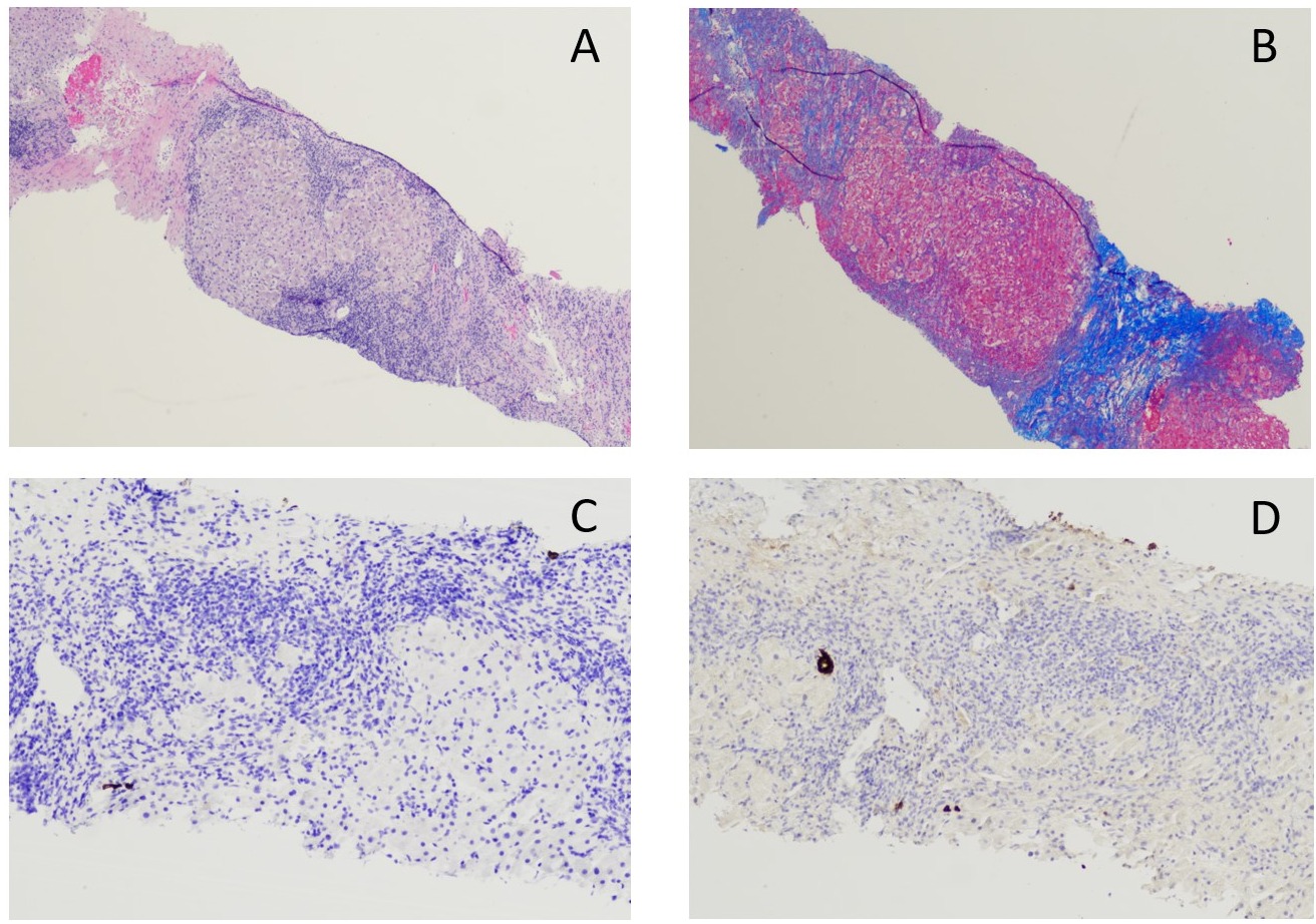
An autoimmune panel was obtained, revealing elevated ANA, pANCA, and dsDNA, with negative AMA and ASMA, low complement level, and normal ceruloplasmin and a-1 antitrypsin. An ultrasound-guided liver biopsy showed cirrhosis with moderate chronic inflammation, and ductopenia with cholestasis as evidenced by minimal scattered cytokeratin 7 and 19 uptake in small bile ducts. Copper deposition was also present. There was no evidence of vasculitis or increased plasma cells.
Discussion: This patient presented with unspecific symptoms, later revealed to be part of a complex autoimmune process causing small-duct cholangiopathy with silent progression to end-stage liver disease. The clinical, immunological, and pathologic findings in this case are not typical of a single condition, suggesting a variant of AIC, containing features of sdPSC and PBC.

Disclosures:
Jose Garcia-Corella, MD, Rupam Sharma, MD, Kasey Fox, DO, Ishaan Kalha, MD. P1375 - Autoimmune Cholangitis: A Rare Case of Silent Liver Cirrhosis, ACG 2024 Annual Scientific Meeting Abstracts. Philadelphia, PA: American College of Gastroenterology.
Kern Medical Center, Bakersfield, CA
Introduction: Autoimmune cholangitis (AIC) is a variant syndrome of autoimmune hepatitis (AIH) which consists of a spectrum of diseases including small-duct primary sclerosing cholangitis (sdPSC), AMA-negative primary biliary cirrhosis (PBC), and IgG4 mediated sclerosing cholangitis.
The immune-mediated bile duct destruction can lead to chronic inflammation and irreversible complications. Here we present a rare case of an AIC variant resulting in cirrhosis in a female patient.
Case Description/Methods: A 41-year-old Hispanic female with no significant medical or social history, presented with one month of progressive abdominal distention, jaundice, peripheral edema, weakness, mild right upper quadrant abdominal pain, and weight loss. Initial workup was notable for abnormal liver function tests (ALT 67 U/L, AST 158 U/L, ALP 332 U/L) and direct hyperbilirubinemia (9.4 mg/dL). Abdominal ultrasound and CT scan showed evidence of cirrhosis with splenomegaly and collateral vessels, suggesting portal venous hypertension with no masses or other signs of obstruction. Noninvasive fibrosis testing showed stage F4 (cirrhosis) with high necrosis inflammatory activity score. EGD showed trace esophageal varices.
An autoimmune panel was obtained, revealing elevated ANA, pANCA, and dsDNA, with negative AMA and ASMA, low complement level, and normal ceruloplasmin and a-1 antitrypsin. An ultrasound-guided liver biopsy showed cirrhosis with moderate chronic inflammation, and ductopenia with cholestasis as evidenced by minimal scattered cytokeratin 7 and 19 uptake in small bile ducts. Copper deposition was also present. There was no evidence of vasculitis or increased plasma cells.
Discussion: This patient presented with unspecific symptoms, later revealed to be part of a complex autoimmune process causing small-duct cholangiopathy with silent progression to end-stage liver disease. The clinical, immunological, and pathologic findings in this case are not typical of a single condition, suggesting a variant of AIC, containing features of sdPSC and PBC.

Figure: Figure 1. Liver biopsy.
A. H&E stain showing nodules surrounded by fibrosis.
B. Trichrome stain showing extensive fibrous bands consistent with advanced cirrhosis.
C & D. Cytokeratin 7 and 19 (respectively) immunostains revealing ductopenia as evident by scattered small duct uptake.
A. H&E stain showing nodules surrounded by fibrosis.
B. Trichrome stain showing extensive fibrous bands consistent with advanced cirrhosis.
C & D. Cytokeratin 7 and 19 (respectively) immunostains revealing ductopenia as evident by scattered small duct uptake.
Disclosures:
Jose Garcia-Corella indicated no relevant financial relationships.
Rupam Sharma indicated no relevant financial relationships.
Kasey Fox indicated no relevant financial relationships.
Ishaan Kalha indicated no relevant financial relationships.
Jose Garcia-Corella, MD, Rupam Sharma, MD, Kasey Fox, DO, Ishaan Kalha, MD. P1375 - Autoimmune Cholangitis: A Rare Case of Silent Liver Cirrhosis, ACG 2024 Annual Scientific Meeting Abstracts. Philadelphia, PA: American College of Gastroenterology.
