Sunday Poster Session
Category: Liver
P1407 - Mysterious Recurrent Ascites Leads to Rare Finding
Sunday, October 27, 2024
3:30 PM - 7:00 PM ET
Location: Exhibit Hall E

Has Audio
- VD
Victoria Diaz, MD
Valley Hospital Medical Center
Las Vegas, NV
Presenting Author(s)
Victoria Diaz, MD, Christina Joya, DO, Masuma Syed, MD, Vishvinder Sharma, MD
Valley Hospital Medical Center, Las Vegas, NV
Introduction: Mesenteric cysts (MC), also called lymphangiomas, are rare defects that present with nonspecific symptoms and are difficult to diagnose preoperatively. The incidence of MC is estimated to be 0.5-1:100,000 adult admissions. MC most commonly originate from the mesentery of the ileum or ascending colon, occurring predominantly in the small intestine or right mesocolic regions. Herein we present a case of a MC originating from the left colon in a patient with recurrent ascites.
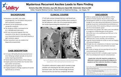
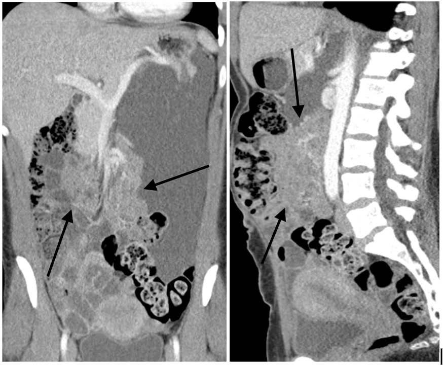
Case Description/Methods: A 31-year-old healthy female presented to our clinic following multiple episodes of recurrent ascites requiring paracentesis. She denied familial history of liver disease. Hepatic panel and synthetic liver function tests were within normal limits. Pelvic ultrasound showed no gynecological abnormalities. CT A/P with contrast showed that her small bowel was largely displaced to the right of midline due to ascites in the left abdomen, with a foci of increased density within the ascites representing a MC. The patient underwent surgery which revealed a large complex cyst involving the left colon and part of the transverse colon. One liter of hemorrhagic fluid was drained and the cyst wall was completely deroofed.
Discussion: While our patient was only 31, the incidence of MC is greatest in females between 40 and 70 years of age. Etiopathogenesis of MC are not fully understood but are thought to occur when abnormal proliferation of mesenteric lymphatic tissues form multiloculated channel dilations on the serosal surface of the mesentery, failing to communicate with the central lymphatic system. The location of our patient’s MC made it even more difficult to recognize as a differential given its left location; 60% of MC occur in the small bowel and 24% occur in the ascending colon. Symptoms are typically non-specific and include chronic abdominal pain, nausea, vomiting, and constipation. Large MC may be mistaken for ascites on exam and imaging, often going undiagnosed. Although very rare, complications of infection, hemorrhage, volvulus, ileus, obstruction and perforation have been observed. MC usually are benign and slow-growing with only 3% of cases presenting with malignancy. Curative treatment for MC is complete surgical resection, often necessitating bowel resection and anastomosis. Despite their rarity, MC should be considered as a differential in patients presenting with ambiguous GI symptoms and fluid collections identified on imaging.

Disclosures:
Victoria Diaz, MD, Christina Joya, DO, Masuma Syed, MD, Vishvinder Sharma, MD. P1407 - Mysterious Recurrent Ascites Leads to Rare Finding, ACG 2024 Annual Scientific Meeting Abstracts. Philadelphia, PA: American College of Gastroenterology.
Valley Hospital Medical Center, Las Vegas, NV
Introduction: Mesenteric cysts (MC), also called lymphangiomas, are rare defects that present with nonspecific symptoms and are difficult to diagnose preoperatively. The incidence of MC is estimated to be 0.5-1:100,000 adult admissions. MC most commonly originate from the mesentery of the ileum or ascending colon, occurring predominantly in the small intestine or right mesocolic regions. Herein we present a case of a MC originating from the left colon in a patient with recurrent ascites.
Case Description/Methods: A 31-year-old healthy female presented to our clinic following multiple episodes of recurrent ascites requiring paracentesis. She denied familial history of liver disease. Hepatic panel and synthetic liver function tests were within normal limits. Pelvic ultrasound showed no gynecological abnormalities. CT A/P with contrast showed that her small bowel was largely displaced to the right of midline due to ascites in the left abdomen, with a foci of increased density within the ascites representing a MC. The patient underwent surgery which revealed a large complex cyst involving the left colon and part of the transverse colon. One liter of hemorrhagic fluid was drained and the cyst wall was completely deroofed.
Discussion: While our patient was only 31, the incidence of MC is greatest in females between 40 and 70 years of age. Etiopathogenesis of MC are not fully understood but are thought to occur when abnormal proliferation of mesenteric lymphatic tissues form multiloculated channel dilations on the serosal surface of the mesentery, failing to communicate with the central lymphatic system. The location of our patient’s MC made it even more difficult to recognize as a differential given its left location; 60% of MC occur in the small bowel and 24% occur in the ascending colon. Symptoms are typically non-specific and include chronic abdominal pain, nausea, vomiting, and constipation. Large MC may be mistaken for ascites on exam and imaging, often going undiagnosed. Although very rare, complications of infection, hemorrhage, volvulus, ileus, obstruction and perforation have been observed. MC usually are benign and slow-growing with only 3% of cases presenting with malignancy. Curative treatment for MC is complete surgical resection, often necessitating bowel resection and anastomosis. Despite their rarity, MC should be considered as a differential in patients presenting with ambiguous GI symptoms and fluid collections identified on imaging.

Figure: CT A/P with contrast showed that small bowel was largely displaced to the right of midline due to large ascites in the left abdomen with a foci of increased density within the ascites favored to represent mesenteric vessels branching from the splenic vein. Arrows pointing at mesenteric cyst within ascites.
Disclosures:
Victoria Diaz indicated no relevant financial relationships.
Christina Joya indicated no relevant financial relationships.
Masuma Syed indicated no relevant financial relationships.
Vishvinder Sharma indicated no relevant financial relationships.
Victoria Diaz, MD, Christina Joya, DO, Masuma Syed, MD, Vishvinder Sharma, MD. P1407 - Mysterious Recurrent Ascites Leads to Rare Finding, ACG 2024 Annual Scientific Meeting Abstracts. Philadelphia, PA: American College of Gastroenterology.
