Sunday Poster Session
Category: Pediatrics
P1482 - A Hidden Culprit: Rare Choledochocyst in a Child With Abdominal Pain
Sunday, October 27, 2024
3:30 PM - 7:00 PM ET
Location: Exhibit Hall E

Has Audio

James Laney, MD
University of Tennessee Health Science Center
Columbia, SC
Presenting Author(s)
James Laney, MD, William Oelsner, MD, Andrew Mims, MD, Samuel Igbinedion, MD
University of Tennessee Health Science Center, Chattanooga, TN
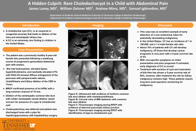
Introduction: A choledochal cyst (CC), is an acquired or congenital anomaly that leads to dilation of intra and extrahepatic portions of the biliary tree. Though the exact etiology of CC remains unclear, the leading theory is that they result from an anomalous pancreaticobiliary junction (APBJ), where the pancreatic and biliary duct join proximal to the sphincter of Oddi. The resultant long channel proximal to the sphincter allows for addition of pancreatic secretions into the common duct which leads to inflammation, dilation, epithelial damage, fibrosis, and cyst formation. We present this rare case of CC in a child presenting with abdominal pain
Case Description/Methods: The patient was a previously healthy 6-year-old female who presented following a weeklong course of progressive generalized abdominal pain with emesis. Labs were notable for lipase of 3,197, t-bili of 1.4, AST of 317, ALT of 682, WBC of 11.2, and Hgb of 14.2. RUQ US showed diffuse enlargement of the pancreas with peripancreatic edema, cholelithiasis and biliary dilation with CBD of 11mm for which GI was consulted for evaluation with ERCP prior to a planned cholecystectomy. Choledocholithiasis was found during ERCP, complete removal was accomplished by biliary sphincterotomy and balloon extraction. Upon review of cholangiogram there was concern for an APBJ, later confirmed on MRCP with a long common channel of 10 mm. Dilation of the extrahepatic common bile duct with milder intrahepatic ductal dilation raised concern for presence of a type Ia choledochal cyst. Cholecystectomy was deferred and patient was discharged home with a plan for hepaticojejunostomy with hepatobiliary surgery
Discussion: A CC is an extremely rare finding in children in the US with an incidence of 1 in 150,000, and a 1:4 male-female rate ratio. About 10% of patients with CC will develop malignancy. Of those that develop cancer, prognosis is very poor with a 5-year survival rate of 5%. This patient had a type Ia CC, a cyst of the extrahepatic bile duct, which accounts for 90% of all CC. The presentation of CC is typically with non-specific symptoms of jaundice, abdominal pain, and palpable abdominal mass. With these non-specific findings on initial presentation and poor prognosis if untreated, early diagnosis of CC is crucial. Treatment with excision of cyst and restoration of bile flow has shown a 5-year survival rate of 90%, however, after treatment the risk for biliary malignancy remains high. These patients require long term post-operative monitoring for malignancy.

Disclosures:
James Laney, MD, William Oelsner, MD, Andrew Mims, MD, Samuel Igbinedion, MD. P1482 - A Hidden Culprit: Rare Choledochocyst in a Child With Abdominal Pain, ACG 2024 Annual Scientific Meeting Abstracts. Philadelphia, PA: American College of Gastroenterology.
University of Tennessee Health Science Center, Chattanooga, TN
Introduction: A choledochal cyst (CC), is an acquired or congenital anomaly that leads to dilation of intra and extrahepatic portions of the biliary tree. Though the exact etiology of CC remains unclear, the leading theory is that they result from an anomalous pancreaticobiliary junction (APBJ), where the pancreatic and biliary duct join proximal to the sphincter of Oddi. The resultant long channel proximal to the sphincter allows for addition of pancreatic secretions into the common duct which leads to inflammation, dilation, epithelial damage, fibrosis, and cyst formation. We present this rare case of CC in a child presenting with abdominal pain
Case Description/Methods: The patient was a previously healthy 6-year-old female who presented following a weeklong course of progressive generalized abdominal pain with emesis. Labs were notable for lipase of 3,197, t-bili of 1.4, AST of 317, ALT of 682, WBC of 11.2, and Hgb of 14.2. RUQ US showed diffuse enlargement of the pancreas with peripancreatic edema, cholelithiasis and biliary dilation with CBD of 11mm for which GI was consulted for evaluation with ERCP prior to a planned cholecystectomy. Choledocholithiasis was found during ERCP, complete removal was accomplished by biliary sphincterotomy and balloon extraction. Upon review of cholangiogram there was concern for an APBJ, later confirmed on MRCP with a long common channel of 10 mm. Dilation of the extrahepatic common bile duct with milder intrahepatic ductal dilation raised concern for presence of a type Ia choledochal cyst. Cholecystectomy was deferred and patient was discharged home with a plan for hepaticojejunostomy with hepatobiliary surgery
Discussion: A CC is an extremely rare finding in children in the US with an incidence of 1 in 150,000, and a 1:4 male-female rate ratio. About 10% of patients with CC will develop malignancy. Of those that develop cancer, prognosis is very poor with a 5-year survival rate of 5%. This patient had a type Ia CC, a cyst of the extrahepatic bile duct, which accounts for 90% of all CC. The presentation of CC is typically with non-specific symptoms of jaundice, abdominal pain, and palpable abdominal mass. With these non-specific findings on initial presentation and poor prognosis if untreated, early diagnosis of CC is crucial. Treatment with excision of cyst and restoration of bile flow has shown a 5-year survival rate of 90%, however, after treatment the risk for biliary malignancy remains high. These patients require long term post-operative monitoring for malignancy.

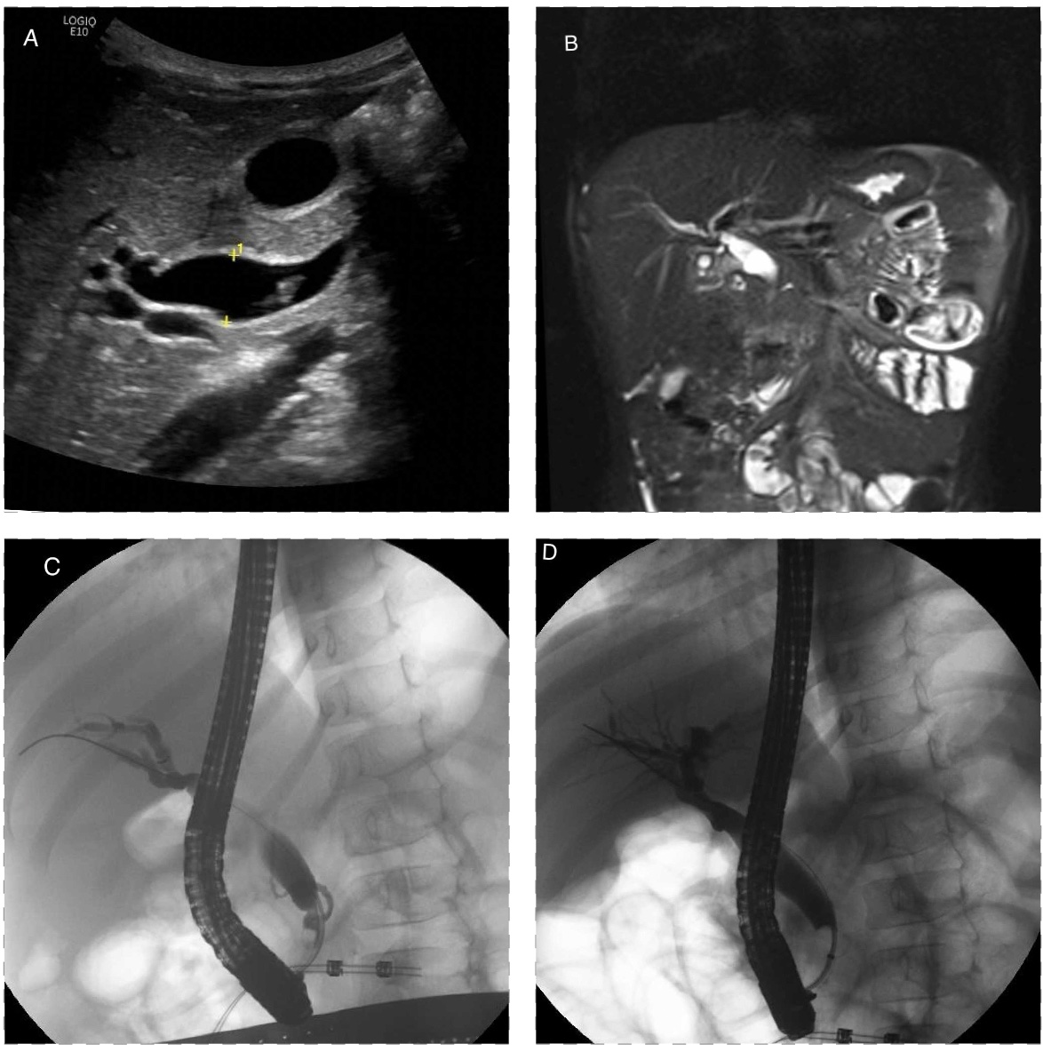
Figure: Figure A: Ultrasound with evidence of fusiform common bile duct dilation with choledocholithiasis. Figure B: Coronal view of MRI abdomen with common bile duct dilation. Figure C: Fluoroscopic imaging during ERCP with evidence of-aberrant pancreatic biliary junction. Figure D: Fluoroscopic imaging during ERCP with identification of type Ia choledochal cyst
Disclosures:
James Laney indicated no relevant financial relationships.
William Oelsner indicated no relevant financial relationships.
Andrew Mims indicated no relevant financial relationships.
Samuel Igbinedion indicated no relevant financial relationships.
James Laney, MD, William Oelsner, MD, Andrew Mims, MD, Samuel Igbinedion, MD. P1482 - A Hidden Culprit: Rare Choledochocyst in a Child With Abdominal Pain, ACG 2024 Annual Scientific Meeting Abstracts. Philadelphia, PA: American College of Gastroenterology.
