Sunday Poster Session
Category: Small Intestine
P1533 - Duodenorenal Fistula Post-Lithotripsy: A Rare Complication
Sunday, October 27, 2024
3:30 PM - 7:00 PM ET
Location: Exhibit Hall E

Has Audio
.jpg)
Anam Zara, MD
HCA Healthcare
Kingwood, TX
Presenting Author(s)
Anam Zara, MD1, Aarohi Parikh, MD1, Ismail Hader, MD2, Duyen Quach, MD1, George Elias, MD1
1HCA Healthcare, Kingwood, TX; 2HCA Kingwood, Kingwood, TX
Introduction: Lithotripsy, though generally safe, can lead to rare complications such as duodenorenal fistula (DRF). DRF is an abnormal connection between the duodenum and kidney, often arising from mechanical trauma such as during lithotripsy.
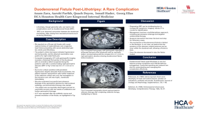
Case Description/Methods: We report a 52-year-old female with a past medical history of nephrolithiasis and congestive heart failure presenting with severe abdominal pain two weeks post-lithotripsy. The patient's initial vital signs indicated hypotension and tachycardia, and the physical examination was positive for right-sided flank pain. Computed tomography (CT) with gastrografin imaging revealed a thickened first portion of the duodenum with an ulcerative defect suspected along the posterior wall with a communicating right perinephric abscess (DRF in Fig-1) that was 8.9 x 4.1 x 8.8 cm in size. Due to her critical condition with persistent hypotension despite adequate fluid resuscitation, the patient required vasopressors with further treatment in the intensive critical care unit. Her hemoglobin in the intensive care unit was 6 g/dL, transfused immediately. She also underwent successful percutaneous drainage of the perinephric abscess by interventional radiology, and antimicrobial therapy was started. The patient was successfully discharged and had an uneventful recovery with two weeks of antibiotics and further perinephric drainage. A CT was repeated after the antibiotic course and showed resolution of the fistula, as highlighted in Fig-2.
Discussion: Diagnosing DRF can be challenging due to nonspecific symptoms. Imaging, mainly CT is crucial for identification. Management involves a multidisciplinary approach, including percutaneous drainage and targeted antimicrobial therapy. Surgical intervention becomes the best next step for refractory cases. The mechanism of DRF is limited in the literature. In our case, we believe that the presence of the abscess created pressure and an ulcer within the duodenal wall, allowing a fistula to form. Once the abscess decreased in size, the fistula healed. DRF post-lithotripsy is rare but requires prompt recognition and interdisciplinary management. Continued reporting and research are vital for understanding and managing this complication effectively.

Disclosures:
Anam Zara, MD1, Aarohi Parikh, MD1, Ismail Hader, MD2, Duyen Quach, MD1, George Elias, MD1. P1533 - Duodenorenal Fistula Post-Lithotripsy: A Rare Complication, ACG 2024 Annual Scientific Meeting Abstracts. Philadelphia, PA: American College of Gastroenterology.
1HCA Healthcare, Kingwood, TX; 2HCA Kingwood, Kingwood, TX
Introduction: Lithotripsy, though generally safe, can lead to rare complications such as duodenorenal fistula (DRF). DRF is an abnormal connection between the duodenum and kidney, often arising from mechanical trauma such as during lithotripsy.
Case Description/Methods: We report a 52-year-old female with a past medical history of nephrolithiasis and congestive heart failure presenting with severe abdominal pain two weeks post-lithotripsy. The patient's initial vital signs indicated hypotension and tachycardia, and the physical examination was positive for right-sided flank pain. Computed tomography (CT) with gastrografin imaging revealed a thickened first portion of the duodenum with an ulcerative defect suspected along the posterior wall with a communicating right perinephric abscess (DRF in Fig-1) that was 8.9 x 4.1 x 8.8 cm in size. Due to her critical condition with persistent hypotension despite adequate fluid resuscitation, the patient required vasopressors with further treatment in the intensive critical care unit. Her hemoglobin in the intensive care unit was 6 g/dL, transfused immediately. She also underwent successful percutaneous drainage of the perinephric abscess by interventional radiology, and antimicrobial therapy was started. The patient was successfully discharged and had an uneventful recovery with two weeks of antibiotics and further perinephric drainage. A CT was repeated after the antibiotic course and showed resolution of the fistula, as highlighted in Fig-2.
Discussion: Diagnosing DRF can be challenging due to nonspecific symptoms. Imaging, mainly CT is crucial for identification. Management involves a multidisciplinary approach, including percutaneous drainage and targeted antimicrobial therapy. Surgical intervention becomes the best next step for refractory cases. The mechanism of DRF is limited in the literature. In our case, we believe that the presence of the abscess created pressure and an ulcer within the duodenal wall, allowing a fistula to form. Once the abscess decreased in size, the fistula healed. DRF post-lithotripsy is rare but requires prompt recognition and interdisciplinary management. Continued reporting and research are vital for understanding and managing this complication effectively.

Figure: Fig-1 Computed tomography with gastrografin highlights a thickened first part of the duodenum with an ulcerative defect, communicating with the complex 8.9 x 4.1 x 8.8 cm, right perinephric abscess showing duodenorenal fistula before treatment. Fig-2 Computed tomography shows post-perinephric abscess drainage after completing antibiotic therapy, highlighting no fistula.
Disclosures:
Anam Zara indicated no relevant financial relationships.
Aarohi Parikh indicated no relevant financial relationships.
Ismail Hader indicated no relevant financial relationships.
Duyen Quach indicated no relevant financial relationships.
George Elias indicated no relevant financial relationships.
Anam Zara, MD1, Aarohi Parikh, MD1, Ismail Hader, MD2, Duyen Quach, MD1, George Elias, MD1. P1533 - Duodenorenal Fistula Post-Lithotripsy: A Rare Complication, ACG 2024 Annual Scientific Meeting Abstracts. Philadelphia, PA: American College of Gastroenterology.
