Sunday Poster Session
Category: Small Intestine
P1563 - A Case of Mycobacterial Duodenitis
Sunday, October 27, 2024
3:30 PM - 7:00 PM ET
Location: Exhibit Hall E

Has Audio

Allison Derise, MD
Louisiana State University Health
Kenner, LA
Presenting Author(s)
Allison Derise, MD1, Hunter Hall, MD2, Stephen Landreneau, MD2
1Louisiana State University, Kenner, LA; 2LSU, New Orleans, LA
Introduction: Mycobacterium avium complex (MAC) is typically an opportunistic infection seen in patients with HIV/AIDS. Involvement of any part of the GI tract can be seen but most commonly affects the duodenum. Appearance on endoscopy has been described most often as white patches. Histopathology can differ among patients with intracellular infiltrates and intensity of acid-fast stains. Of the cases reported, symptoms are typically vague, so disseminated MAC must always remain on the differential in immunocompromised patients. We present a case of mycobacterium duodenitis presenting as symptomatic anemia and concern for shock.
Case Description/Methods: We present a 54-year-old man with a history of AIDS, chronic adrenal insufficiency, disseminated histoplasmosis, resistant disseminated MAC, DLBCL, cryptococcal meningitis, HCV s/p Epclusa who had recent prolonged hospitalization for bacteremia and discharged home with plans for outpatient infusions. He represented for two episodes of black, loose stools and generalized malaise. He does not take blood thinners and has not used OTC medications such as NSAIDs.
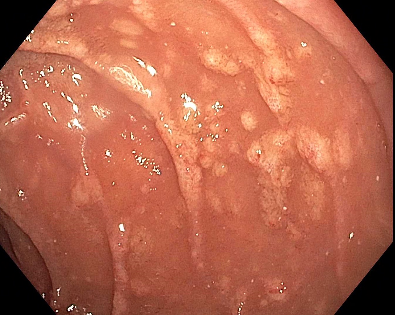
EGD showed shallow esophageal ulcers and abnormal-appearing mucosa to the 2nd portion of the duodenum with biopsies taken in the esophagus, stomach, and duodenum. Small bowel biopsies showed evidence of mycobacterial duodenitis with AFB and PAS statins positive.
He is currently being treated with rifabutin, ethambutol due to known fluroquinolone resistance, and amikacin for resistant disseminated MAC.
Discussion: MAC infection of the GI tract demonstrates a large range of clinical, endoscopic, and histopathological presentations and therefore must always be considered in immunocompromised patients with HIV/AIDS. Biopsies with careful examination under the microscope is essential in correct diagnosis. Treatment is usually a lifelong multidrug regimen with emphasis on adherence to anti-retroviral therapy.

Disclosures:
Allison Derise, MD1, Hunter Hall, MD2, Stephen Landreneau, MD2. P1563 - A Case of Mycobacterial Duodenitis, ACG 2024 Annual Scientific Meeting Abstracts. Philadelphia, PA: American College of Gastroenterology.
1Louisiana State University, Kenner, LA; 2LSU, New Orleans, LA
Introduction: Mycobacterium avium complex (MAC) is typically an opportunistic infection seen in patients with HIV/AIDS. Involvement of any part of the GI tract can be seen but most commonly affects the duodenum. Appearance on endoscopy has been described most often as white patches. Histopathology can differ among patients with intracellular infiltrates and intensity of acid-fast stains. Of the cases reported, symptoms are typically vague, so disseminated MAC must always remain on the differential in immunocompromised patients. We present a case of mycobacterium duodenitis presenting as symptomatic anemia and concern for shock.
Case Description/Methods: We present a 54-year-old man with a history of AIDS, chronic adrenal insufficiency, disseminated histoplasmosis, resistant disseminated MAC, DLBCL, cryptococcal meningitis, HCV s/p Epclusa who had recent prolonged hospitalization for bacteremia and discharged home with plans for outpatient infusions. He represented for two episodes of black, loose stools and generalized malaise. He does not take blood thinners and has not used OTC medications such as NSAIDs.
EGD showed shallow esophageal ulcers and abnormal-appearing mucosa to the 2nd portion of the duodenum with biopsies taken in the esophagus, stomach, and duodenum. Small bowel biopsies showed evidence of mycobacterial duodenitis with AFB and PAS statins positive.
He is currently being treated with rifabutin, ethambutol due to known fluroquinolone resistance, and amikacin for resistant disseminated MAC.
Discussion: MAC infection of the GI tract demonstrates a large range of clinical, endoscopic, and histopathological presentations and therefore must always be considered in immunocompromised patients with HIV/AIDS. Biopsies with careful examination under the microscope is essential in correct diagnosis. Treatment is usually a lifelong multidrug regimen with emphasis on adherence to anti-retroviral therapy.

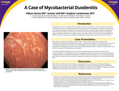
Figure: EGD picture of the 2nd portion of the duodenum showing abnormal mucosa with evidence of white patches that can seen in mycobacterial duodenitis.
Disclosures:
Allison Derise indicated no relevant financial relationships.
Hunter Hall indicated no relevant financial relationships.
Stephen Landreneau indicated no relevant financial relationships.
Allison Derise, MD1, Hunter Hall, MD2, Stephen Landreneau, MD2. P1563 - A Case of Mycobacterial Duodenitis, ACG 2024 Annual Scientific Meeting Abstracts. Philadelphia, PA: American College of Gastroenterology.
