Sunday Poster Session
Category: Small Intestine
P1585 - Ileal Neuroendocrine Tumor: A Rare Incidental Finding
Sunday, October 27, 2024
3:30 PM - 7:00 PM ET
Location: Exhibit Hall E

Has Audio

Abdulmalik Saleem, MD
Henry Ford Health
Flushing, MI
Presenting Author(s)
Abdulmalik Saleem, MD1, Ammad Javaid. Chaudhary, MD1, Taher Jamali, MD2, Momin Samad, MD3, Kimberly Tosch, MD1
1Henry Ford Health, Detroit, MI; 2Henry Ford Hospital, Detroit, MI; 3Henry Ford Hospital, Rochester Hills, MI
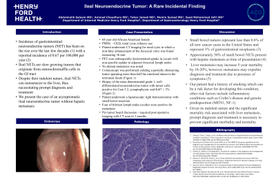
Introduction: Ileal neuroendocrine tumors (NET) are rare, slow growing tumors that originate from enterochromaffin cells in the GI tract. The incidence of these tumors has been on the rise over the last few decades. Patients with small bowel NETs may initially be asymptomatic or may experience vague symptoms. Despite their indolent nature, these tumors may metastasize to the liver necessitating prompt diagnosis and treatment. We present the case of a rare, ileal neuroendocrine tumor discovered as an incidental finding.
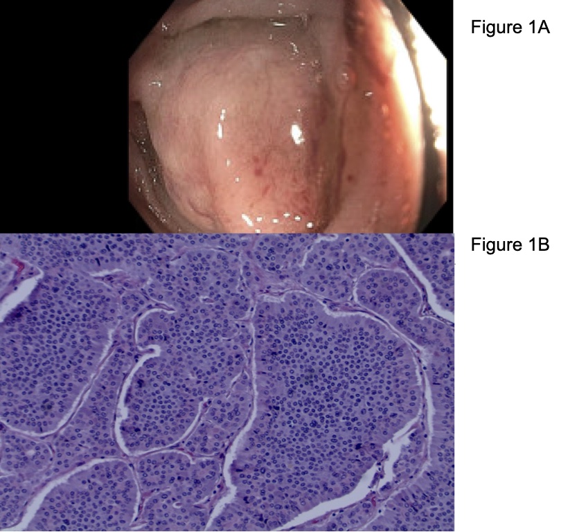
Case Description/Methods: We present the case of an asymptomatic 68-year-old African American male with a history of chronic kidney disease and tobacco use who underwent magnetic resonance imaging of the abdomen for evaluation of renal cysts. Imaging demonstrated complex renal cysts in addition to an incidental finding of a non-fatty enhancement of the ileocecal valve measuring 36 mm. PET scan demonstrated increased uptake in the cecum corresponding to the lesion along with non-specific uptake in adjacent ileocecal lymph nodes with no distant metastatic disease noted. Subsequent colonoscopy identified a partially obstructing tumor spanning more than half the intestinal lumen in the terminal ileum (Figure 1A). The mass was biopsied, and pathology revealed a grade 1, well differentiated neuroendocrine tumor with tumor cells positive for Cam 5.2, synaptophysin, and Ki67 < 3% (Figure 1B). A laparoscopic right hemicolectomy with small bowel resection was performed. Four out of thirteen excised adjacent lymph nodes were positive for metastatic neuroendocrine tumor. Our patient’s postoperative course was complicated by ileus with eventual resolution. The case was discussed in a multidisciplinary tumor board and the decision for surveillance alone was made with a repeat CT scan in 3 months.
Discussion: Small bowel tumors represent less than 0.6% of all cancers with neuroendocrine tumors being the most common in this subset. More than half of these patients present with liver metastasis at the time of diagnosis. Liver metastasis may increase 5-year mortality by 10 – 20%, however, patients with liver metastasis are more likely to be symptomatic expediting diagnosis. Clinicians should be wary of certain risk factors predisposing patients to this pathology such as smoking and the presence of Crohn’s disease or genetic syndromes such as MEN1. Given its indolent nature and the mortality associated with liver metastasis, prompt diagnosis and treatment is necessary in these patients.

Disclosures:
Abdulmalik Saleem, MD1, Ammad Javaid. Chaudhary, MD1, Taher Jamali, MD2, Momin Samad, MD3, Kimberly Tosch, MD1. P1585 - Ileal Neuroendocrine Tumor: A Rare Incidental Finding, ACG 2024 Annual Scientific Meeting Abstracts. Philadelphia, PA: American College of Gastroenterology.
1Henry Ford Health, Detroit, MI; 2Henry Ford Hospital, Detroit, MI; 3Henry Ford Hospital, Rochester Hills, MI
Introduction: Ileal neuroendocrine tumors (NET) are rare, slow growing tumors that originate from enterochromaffin cells in the GI tract. The incidence of these tumors has been on the rise over the last few decades. Patients with small bowel NETs may initially be asymptomatic or may experience vague symptoms. Despite their indolent nature, these tumors may metastasize to the liver necessitating prompt diagnosis and treatment. We present the case of a rare, ileal neuroendocrine tumor discovered as an incidental finding.
Case Description/Methods: We present the case of an asymptomatic 68-year-old African American male with a history of chronic kidney disease and tobacco use who underwent magnetic resonance imaging of the abdomen for evaluation of renal cysts. Imaging demonstrated complex renal cysts in addition to an incidental finding of a non-fatty enhancement of the ileocecal valve measuring 36 mm. PET scan demonstrated increased uptake in the cecum corresponding to the lesion along with non-specific uptake in adjacent ileocecal lymph nodes with no distant metastatic disease noted. Subsequent colonoscopy identified a partially obstructing tumor spanning more than half the intestinal lumen in the terminal ileum (Figure 1A). The mass was biopsied, and pathology revealed a grade 1, well differentiated neuroendocrine tumor with tumor cells positive for Cam 5.2, synaptophysin, and Ki67 < 3% (Figure 1B). A laparoscopic right hemicolectomy with small bowel resection was performed. Four out of thirteen excised adjacent lymph nodes were positive for metastatic neuroendocrine tumor. Our patient’s postoperative course was complicated by ileus with eventual resolution. The case was discussed in a multidisciplinary tumor board and the decision for surveillance alone was made with a repeat CT scan in 3 months.
Discussion: Small bowel tumors represent less than 0.6% of all cancers with neuroendocrine tumors being the most common in this subset. More than half of these patients present with liver metastasis at the time of diagnosis. Liver metastasis may increase 5-year mortality by 10 – 20%, however, patients with liver metastasis are more likely to be symptomatic expediting diagnosis. Clinicians should be wary of certain risk factors predisposing patients to this pathology such as smoking and the presence of Crohn’s disease or genetic syndromes such as MEN1. Given its indolent nature and the mortality associated with liver metastasis, prompt diagnosis and treatment is necessary in these patients.

Figure: Figure 1A: partially obstructing tumor in the terminal Ileum on colonoscopy
Figure 2A: pathology, well differentiated neuroendocrine tumor
Figure 2A: pathology, well differentiated neuroendocrine tumor
Disclosures:
Abdulmalik Saleem indicated no relevant financial relationships.
Ammad Chaudhary indicated no relevant financial relationships.
Taher Jamali indicated no relevant financial relationships.
Momin Samad indicated no relevant financial relationships.
Kimberly Tosch indicated no relevant financial relationships.
Abdulmalik Saleem, MD1, Ammad Javaid. Chaudhary, MD1, Taher Jamali, MD2, Momin Samad, MD3, Kimberly Tosch, MD1. P1585 - Ileal Neuroendocrine Tumor: A Rare Incidental Finding, ACG 2024 Annual Scientific Meeting Abstracts. Philadelphia, PA: American College of Gastroenterology.
