Sunday Poster Session
Category: Small Intestine
P1586 - Uncommon Manifestation of Gallstone Ileus in a Middle-Aged Male
Sunday, October 27, 2024
3:30 PM - 7:00 PM ET
Location: Exhibit Hall E

Has Audio

Cynthia D. Varela
St. George's University School of Medicine
Hemet, CA
Presenting Author(s)
Cynthia D. Varela, 1, Daniel Igwe, MD2
1St. George's University School of Medicine, Hemet, CA; 2Hemet and Menifee Global Medical Centers, Hemet, CA
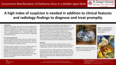
Introduction: Gallstone ileus has been found in 0.3% to 0.5% of patients with gallstones. Most patients are female, in a ratio of 3.5 females to 1 male, and commonly over the age of 60. It is more rare to have mechanical obstructions (less than 0.1%) from a gallstone ileus due to a tumbling gallstone from a cholecystoduodenal fistula with pneumobilia. We present a case occuring in a middle-aged male with liver disease.
Case Description/Methods: A 52-year-old bed-bound male with a history of 2 strokes with left sided hemiplegia and alcoholic cirrhosis presented with “stabbing” upper abdominal pain for 2 weeks. When amber-colored vomit was seen by his caregiver he was taken to the ER.
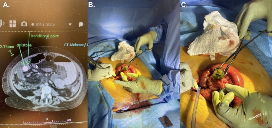
CT Abdomen/Pelvis revealed a 21 mm x 21 mm gallstone that had eroded through the gallbladder into the first portion of the duodenum with small amounts of air in the biliary tree and in the gallbladder. Patient was subsequently diagnosed gallstone ileus within 48 hours of admission secondary to a tumbling stone through a cholecystoduodenal fistula associated with pneumobilia. The stone in the duodenum did not initially present with obstructive symptoms.
After the noted erosion of the gallstone into the duodenum, the patient developed obstructive symptoms within 24-48 hours consisting of obstipation, progressive abdominal distention, and frank gallstone ileus. Subsequent CT showed a tumbling stone with ileus. Patient was taken to the OR for an open procedure with an enterotomy of a 3.5 cm stone extracted 2 feet from the ileocecal valve. The small bowel was ran through to rule out more tumbling stones and the gallbladder could not be well palpated for stones due to omental adhesions and cirrhotic liver. The patient was admitted to ICU for closer monitoring, he tolerated the procedure well. Patient was fed on post-op day 3 then was advanced as tolerated prior to discharge.
Discussion: The treatment for gallstone ileus is emergent surgery to reduce complications. This patient did have nonspecific symptoms therefore imaging played a big part in detection leading to proper treatment.
According to Turner et al, “high morbidity and mortality rates have been reported for patients with gallstone ileus.” The exact treatment of gallstone ileus is controversial, but the main goal is removal of the stone as was done in this case. A high index of suspicion is needed in addition to clinical features and radiology findings to diagnose and treat promptly.

Disclosures:
Cynthia D. Varela, 1, Daniel Igwe, MD2. P1586 - Uncommon Manifestation of Gallstone Ileus in a Middle-Aged Male, ACG 2024 Annual Scientific Meeting Abstracts. Philadelphia, PA: American College of Gastroenterology.
1St. George's University School of Medicine, Hemet, CA; 2Hemet and Menifee Global Medical Centers, Hemet, CA
Introduction: Gallstone ileus has been found in 0.3% to 0.5% of patients with gallstones. Most patients are female, in a ratio of 3.5 females to 1 male, and commonly over the age of 60. It is more rare to have mechanical obstructions (less than 0.1%) from a gallstone ileus due to a tumbling gallstone from a cholecystoduodenal fistula with pneumobilia. We present a case occuring in a middle-aged male with liver disease.
Case Description/Methods: A 52-year-old bed-bound male with a history of 2 strokes with left sided hemiplegia and alcoholic cirrhosis presented with “stabbing” upper abdominal pain for 2 weeks. When amber-colored vomit was seen by his caregiver he was taken to the ER.
CT Abdomen/Pelvis revealed a 21 mm x 21 mm gallstone that had eroded through the gallbladder into the first portion of the duodenum with small amounts of air in the biliary tree and in the gallbladder. Patient was subsequently diagnosed gallstone ileus within 48 hours of admission secondary to a tumbling stone through a cholecystoduodenal fistula associated with pneumobilia. The stone in the duodenum did not initially present with obstructive symptoms.
After the noted erosion of the gallstone into the duodenum, the patient developed obstructive symptoms within 24-48 hours consisting of obstipation, progressive abdominal distention, and frank gallstone ileus. Subsequent CT showed a tumbling stone with ileus. Patient was taken to the OR for an open procedure with an enterotomy of a 3.5 cm stone extracted 2 feet from the ileocecal valve. The small bowel was ran through to rule out more tumbling stones and the gallbladder could not be well palpated for stones due to omental adhesions and cirrhotic liver. The patient was admitted to ICU for closer monitoring, he tolerated the procedure well. Patient was fed on post-op day 3 then was advanced as tolerated prior to discharge.
Discussion: The treatment for gallstone ileus is emergent surgery to reduce complications. This patient did have nonspecific symptoms therefore imaging played a big part in detection leading to proper treatment.
According to Turner et al, “high morbidity and mortality rates have been reported for patients with gallstone ileus.” The exact treatment of gallstone ileus is controversial, but the main goal is removal of the stone as was done in this case. A high index of suspicion is needed in addition to clinical features and radiology findings to diagnose and treat promptly.

Figure: A. CT Abd/Pelvis shows a migrated gallstone at the transition point of the ileocecal valve.
B. & C. Show an extracted 3.5 cm stone during enterotomy 2 feet from the ileocecal valve.
B. & C. Show an extracted 3.5 cm stone during enterotomy 2 feet from the ileocecal valve.
Disclosures:
Cynthia Varela indicated no relevant financial relationships.
Daniel Igwe indicated no relevant financial relationships.
Cynthia D. Varela, 1, Daniel Igwe, MD2. P1586 - Uncommon Manifestation of Gallstone Ileus in a Middle-Aged Male, ACG 2024 Annual Scientific Meeting Abstracts. Philadelphia, PA: American College of Gastroenterology.
