Sunday Poster Session
Category: Stomach
P1634 - Isolated Gastric Varices in the Setting of Left-Sided Portal Hypertension: A Very Rare Cause of Gastrointestinal Bleeding
Sunday, October 27, 2024
3:30 PM - 7:00 PM ET
Location: Exhibit Hall E

Has Audio

Yasir Ahmed, MD
United Health Services
Binghamton, NY
Presenting Author(s)
Yasir Ahmed, MD1, Usama Sakhawat, MD2, Ibrar Atiq, MD2, Gowthami Ramar, MD1, Fahad Malik, MD2, Toseef Javaid, MD2
1United Health Services, Binghamton, NY; 2United Health Services, Wilson Medical Center, Binghamton, NY
Introduction: Isolated left-sided portal hypertension (ILPH) is a rare clinical syndrome caused by splenic vein obstruction or inadequate drainage. It is also called sinistral portal hypertension. Pancreatitis, pancreatic neoplasm are relatively common etiologies of splenic vein obstruction, while pancreatic pseudocyst, prior pancreatic surgery, gastric, renal, or retroperitoneal pathology, arterial pseudoaneurysm, tuberculous adenitis, or thrombocythemia are the less common etiologies.
Case Description/Methods:
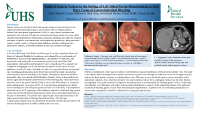
A 67-year-old female with diabetes mellitus type II, anxiety, hypothyroidism and history of classical Hodgkin’s lymphoma was evaluated for several episodes of hematemesis and associated lightheadedness. The patient was tremulous, and hypotensive and tachycardic on presentation and received immediate fluid resuscitation. Hemoglobin and hematocrit were 6.3 mg/dL and 19.2, respectively. Computed tomography scan of the abdomen and pelvis did not show an acute pathology. A total of 5 units of packed red cells were transfused during the hospital stay. Esophagogastroduodenoscopy (EGD) showed red blood and one spurting cratered gastric ulcer hemorrhage in the fundus. Epinephrine injections and three hemostatic clips were placed and the bleeding stopped. A drop in hemoglobin was noticed again and the patient underwent EGD the following day, which showed an ulcerated area in the gastric fundus (likely a varix) with fibrin plug in it, consistent with isolated gastric varix type I (IGV-1). A second linear ulcerated area without active bleeding was seen along the greater curvature as well (likely a decompressed ulcerated varix). A CT angiogram of the abdomen and pelvis confirmed large gastric varices due to left-sided portal hypertension, likely due to distal pancreatectomy in the past vs thrombosis. The patient had a laparoscopic distal pancreatectomy with spleen preservation done for benign ducal tumor done >10 years ago.
A laparoscopic splenectomy was performed, the patient tolerated the procedure well and was discharged home in stable condition after recovery.
Discussion: Extrinsic compression of any cause or splenic vein thrombosis causes hypertension of the proximal splenic vein, leading to collateral drainage and varices formation. Splenic arteriography is the gold standard for diagnosis and splenectomy is recommended for bleeding gastric varices.
Disclosures:
Yasir Ahmed, MD1, Usama Sakhawat, MD2, Ibrar Atiq, MD2, Gowthami Ramar, MD1, Fahad Malik, MD2, Toseef Javaid, MD2. P1634 - Isolated Gastric Varices in the Setting of Left-Sided Portal Hypertension: A Very Rare Cause of Gastrointestinal Bleeding, ACG 2024 Annual Scientific Meeting Abstracts. Philadelphia, PA: American College of Gastroenterology.
1United Health Services, Binghamton, NY; 2United Health Services, Wilson Medical Center, Binghamton, NY
Introduction: Isolated left-sided portal hypertension (ILPH) is a rare clinical syndrome caused by splenic vein obstruction or inadequate drainage. It is also called sinistral portal hypertension. Pancreatitis, pancreatic neoplasm are relatively common etiologies of splenic vein obstruction, while pancreatic pseudocyst, prior pancreatic surgery, gastric, renal, or retroperitoneal pathology, arterial pseudoaneurysm, tuberculous adenitis, or thrombocythemia are the less common etiologies.
Case Description/Methods:
A 67-year-old female with diabetes mellitus type II, anxiety, hypothyroidism and history of classical Hodgkin’s lymphoma was evaluated for several episodes of hematemesis and associated lightheadedness. The patient was tremulous, and hypotensive and tachycardic on presentation and received immediate fluid resuscitation. Hemoglobin and hematocrit were 6.3 mg/dL and 19.2, respectively. Computed tomography scan of the abdomen and pelvis did not show an acute pathology. A total of 5 units of packed red cells were transfused during the hospital stay. Esophagogastroduodenoscopy (EGD) showed red blood and one spurting cratered gastric ulcer hemorrhage in the fundus. Epinephrine injections and three hemostatic clips were placed and the bleeding stopped. A drop in hemoglobin was noticed again and the patient underwent EGD the following day, which showed an ulcerated area in the gastric fundus (likely a varix) with fibrin plug in it, consistent with isolated gastric varix type I (IGV-1). A second linear ulcerated area without active bleeding was seen along the greater curvature as well (likely a decompressed ulcerated varix). A CT angiogram of the abdomen and pelvis confirmed large gastric varices due to left-sided portal hypertension, likely due to distal pancreatectomy in the past vs thrombosis. The patient had a laparoscopic distal pancreatectomy with spleen preservation done for benign ducal tumor done >10 years ago.
A laparoscopic splenectomy was performed, the patient tolerated the procedure well and was discharged home in stable condition after recovery.
Discussion: Extrinsic compression of any cause or splenic vein thrombosis causes hypertension of the proximal splenic vein, leading to collateral drainage and varices formation. Splenic arteriography is the gold standard for diagnosis and splenectomy is recommended for bleeding gastric varices.
Disclosures:
Yasir Ahmed indicated no relevant financial relationships.
Usama Sakhawat indicated no relevant financial relationships.
Ibrar Atiq indicated no relevant financial relationships.
Gowthami Ramar indicated no relevant financial relationships.
Fahad Malik indicated no relevant financial relationships.
Toseef Javaid indicated no relevant financial relationships.
Yasir Ahmed, MD1, Usama Sakhawat, MD2, Ibrar Atiq, MD2, Gowthami Ramar, MD1, Fahad Malik, MD2, Toseef Javaid, MD2. P1634 - Isolated Gastric Varices in the Setting of Left-Sided Portal Hypertension: A Very Rare Cause of Gastrointestinal Bleeding, ACG 2024 Annual Scientific Meeting Abstracts. Philadelphia, PA: American College of Gastroenterology.

