Sunday Poster Session
Category: Stomach
P1654 - Hidden in Plain Sight: Foreign Body Ingestion
Sunday, October 27, 2024
3:30 PM - 7:00 PM ET
Location: Exhibit Hall E

Has Audio

Allison Derise, MD
Louisiana State University Health
Kenner, LA
Presenting Author(s)
Allison Derise, MD1, Catherine T.. Hudson, MD, MPH2
1Louisiana State University Health, Kenner, LA; 2LSU, New Orleans, LA
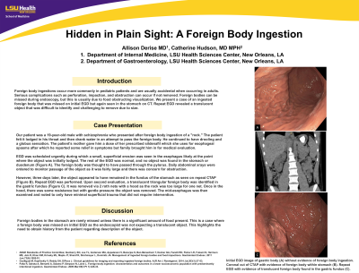
Introduction: Foreign body ingestions occur more commonly in pediatric patients and are usually accidental when occurring in adults. Serious complications such as perforation, impaction, and obstruction can occur if not removed. Foreign bodies can be missed during endoscopy, but this is usually due to food obstructing visualization. We present a case of an ingested foreign body that was missed on initial EGD but again seen in the stomach on CT. Repeat EGD revealed a translucent object that was difficult to identify and challenging to remove due to size.
Case Description/Methods: Our patient was a 19-year-old male with schizophrenia who presented after foreign body ingestion of a "rock." The patient felt it lodged in his throat and then drank water in an attempt to pass the foreign body. He continued to have drooling and a globus sensation. The patient’s mother gave him a dose of her prescribed sildenafil which she uses for esophageal spasms after which he reported some relief in symptoms but family brought him in for medical evaluation.
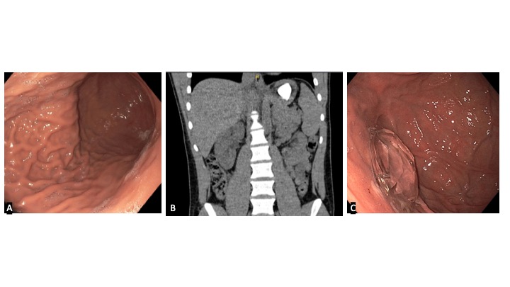
EGD was scheduled urgently during which a small, superficial erosion was seen in the esophagus likely at the point where the object was initially lodged. The rest of the EGD was normal, and no object was found in the stomach or duodenum (Figure A). The foreign body was thought to have passed through the pylorus. Daily abdominal xrays were ordered to monitor passage of the object as it was fairly large and there was concern for obstruction.
However, three days later, the object appeared to have remained in the fundus of the stomach as seen on repeat CTAP (Figure B). Repeat EGD was performed. Upon second evaluation, a translucent triangular foreign body was identified in the gastric fundus (Figure C). It was removed via 2 roth nets with a hood as the rock was too large for one net. Once in the hood, there was some resistance but with gentle pressure the object was removed. The mid-esophagus was then examined and noted to only have minimal superficial trauma that did not require intervention.
Discussion: Foreign bodies in the stomach are rarely missed unless there is a significant amount of food present. This is a case where a foreign body was missed on initial EGD as the endoscopist was not expecting a translucent object. This highlights the need to obtain history from the patient regarding description of the object.

Disclosures:
Allison Derise, MD1, Catherine T.. Hudson, MD, MPH2. P1654 - Hidden in Plain Sight: Foreign Body Ingestion, ACG 2024 Annual Scientific Meeting Abstracts. Philadelphia, PA: American College of Gastroenterology.
1Louisiana State University Health, Kenner, LA; 2LSU, New Orleans, LA
Introduction: Foreign body ingestions occur more commonly in pediatric patients and are usually accidental when occurring in adults. Serious complications such as perforation, impaction, and obstruction can occur if not removed. Foreign bodies can be missed during endoscopy, but this is usually due to food obstructing visualization. We present a case of an ingested foreign body that was missed on initial EGD but again seen in the stomach on CT. Repeat EGD revealed a translucent object that was difficult to identify and challenging to remove due to size.
Case Description/Methods: Our patient was a 19-year-old male with schizophrenia who presented after foreign body ingestion of a "rock." The patient felt it lodged in his throat and then drank water in an attempt to pass the foreign body. He continued to have drooling and a globus sensation. The patient’s mother gave him a dose of her prescribed sildenafil which she uses for esophageal spasms after which he reported some relief in symptoms but family brought him in for medical evaluation.
EGD was scheduled urgently during which a small, superficial erosion was seen in the esophagus likely at the point where the object was initially lodged. The rest of the EGD was normal, and no object was found in the stomach or duodenum (Figure A). The foreign body was thought to have passed through the pylorus. Daily abdominal xrays were ordered to monitor passage of the object as it was fairly large and there was concern for obstruction.
However, three days later, the object appeared to have remained in the fundus of the stomach as seen on repeat CTAP (Figure B). Repeat EGD was performed. Upon second evaluation, a translucent triangular foreign body was identified in the gastric fundus (Figure C). It was removed via 2 roth nets with a hood as the rock was too large for one net. Once in the hood, there was some resistance but with gentle pressure the object was removed. The mid-esophagus was then examined and noted to only have minimal superficial trauma that did not require intervention.
Discussion: Foreign bodies in the stomach are rarely missed unless there is a significant amount of food present. This is a case where a foreign body was missed on initial EGD as the endoscopist was not expecting a translucent object. This highlights the need to obtain history from the patient regarding description of the object.

Figure: Initial EGD image of gastric body (A) without evidence of foreign body ingestion. Coronal cut of CTAP with evidence of foreign body within stomach (B). Repeat EGD with evidence of translucent foreign body found in the gastric fundus (C).
Disclosures:
Allison Derise indicated no relevant financial relationships.
Catherine Hudson indicated no relevant financial relationships.
Allison Derise, MD1, Catherine T.. Hudson, MD, MPH2. P1654 - Hidden in Plain Sight: Foreign Body Ingestion, ACG 2024 Annual Scientific Meeting Abstracts. Philadelphia, PA: American College of Gastroenterology.
