Sunday Poster Session
Category: Stomach
P1658 - Linitis Plastica – An Ancient Scourge Lurking Beneath the Surface
Sunday, October 27, 2024
3:30 PM - 7:00 PM ET
Location: Exhibit Hall E

Has Audio

Amro Abdellatief, MD
Howard University Hospital
Silver Spring, MD
Presenting Author(s)
Amro Abdellatief, MD1, Oluwatayo J. Awolumate, MD2, Sneha Adidam, MD2, Farshad Aduli, MD2, Adeyinka O. Laiyemo, MD, MPH2
1Howard University Hospital, Silver Spring, MD; 2Howard University Hospital, Washington, DC
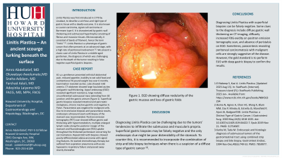
Introduction: Linitis Plastica, also known as Lauren carcinoma, signet cell carcinoma or Borrmann type 4, is characterized by gastric wall thickening and submucosal hypertrophy consisting of fibrous and muscular tissues. Microscopically, it consisted of bands of filament, hence the term “linitis”. [1]. Linitis Plastica often presents at an advanced stage, with a high rate of peritoneal involvement [2]. We present a classic case of Linitis Plastica in a middle-aged gentleman, the diagnosis of which was challenging due to the depth of the tumor resulting in false negative superficial gastric biopsies.
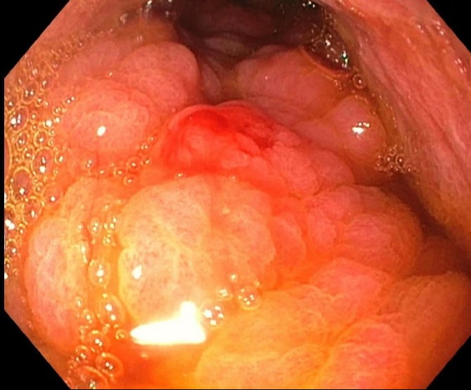
Case Description/Methods: 61 y.o. gentleman presented with dull abdominal pain, reduced appetite, inability to eat solid food and unintentional 70-pound weight loss over 6 months. Examination revealed ascites. Labs showed mild anemia. CT-abdomen showed large loculated ascites and gastric wall thickening. Upper endoscopy (EGD) revealed significant resistance, large nodular, circumferential submucosal mass extending from GE junction until the gastric antrum. (Figure 1 + 2). Superficial gastric biopsies revealed intestinal and pancreatic metaplasia, chronic inactive gastritis and negative H. Pylori. Pt also underwent paracentesis which was negative for malignant cells. Due to inconclusive biopsies and paracentesis results, endoscopic ultrasound exam with biopsy of the stomach was recommended. Positron emission tomography (PET) scan showed diffuse gastric wall thickening with hypermetabolism, multiple enlarged lymph nodes along the posterior margin of the stomach and fluorodeoxyglucose (FDG) uptake throughout the thickened peritoneum concerning for carcinomatosis. Repeat EGD with biopsies showed poorly-differentiated adenocarcinoma with signet ring features. He started chemotherapy therapy but suffered from aspiration pneumonia with acute hypoxemic respiratory failure and passed away shortly thereafter.
Discussion: Diagnosing Linitis Plastica can be challenging due to infiltration of the submucosa and muscularis propria. Superficial gastric biopsies tend to be falsely negative. Therefore, it is recommended to incorporate the combination of strip and bite biopsy techniques when there is a suspicion of a diffuse type of gastric cancer [3]. Some clues to the diagnosis include diffuse gastric wall thickening on CT-imaging, increased FDG-avidity on positron emission tomography scan, and absence of peristalsis on EGD. However, the gold standard is to perform EUS with deep gastric biopsy to confirm the results.

Disclosures:
Amro Abdellatief, MD1, Oluwatayo J. Awolumate, MD2, Sneha Adidam, MD2, Farshad Aduli, MD2, Adeyinka O. Laiyemo, MD, MPH2. P1658 - Linitis Plastica – An Ancient Scourge Lurking Beneath the Surface, ACG 2024 Annual Scientific Meeting Abstracts. Philadelphia, PA: American College of Gastroenterology.
1Howard University Hospital, Silver Spring, MD; 2Howard University Hospital, Washington, DC
Introduction: Linitis Plastica, also known as Lauren carcinoma, signet cell carcinoma or Borrmann type 4, is characterized by gastric wall thickening and submucosal hypertrophy consisting of fibrous and muscular tissues. Microscopically, it consisted of bands of filament, hence the term “linitis”. [1]. Linitis Plastica often presents at an advanced stage, with a high rate of peritoneal involvement [2]. We present a classic case of Linitis Plastica in a middle-aged gentleman, the diagnosis of which was challenging due to the depth of the tumor resulting in false negative superficial gastric biopsies.
Case Description/Methods: 61 y.o. gentleman presented with dull abdominal pain, reduced appetite, inability to eat solid food and unintentional 70-pound weight loss over 6 months. Examination revealed ascites. Labs showed mild anemia. CT-abdomen showed large loculated ascites and gastric wall thickening. Upper endoscopy (EGD) revealed significant resistance, large nodular, circumferential submucosal mass extending from GE junction until the gastric antrum. (Figure 1 + 2). Superficial gastric biopsies revealed intestinal and pancreatic metaplasia, chronic inactive gastritis and negative H. Pylori. Pt also underwent paracentesis which was negative for malignant cells. Due to inconclusive biopsies and paracentesis results, endoscopic ultrasound exam with biopsy of the stomach was recommended. Positron emission tomography (PET) scan showed diffuse gastric wall thickening with hypermetabolism, multiple enlarged lymph nodes along the posterior margin of the stomach and fluorodeoxyglucose (FDG) uptake throughout the thickened peritoneum concerning for carcinomatosis. Repeat EGD with biopsies showed poorly-differentiated adenocarcinoma with signet ring features. He started chemotherapy therapy but suffered from aspiration pneumonia with acute hypoxemic respiratory failure and passed away shortly thereafter.
Discussion: Diagnosing Linitis Plastica can be challenging due to infiltration of the submucosa and muscularis propria. Superficial gastric biopsies tend to be falsely negative. Therefore, it is recommended to incorporate the combination of strip and bite biopsy techniques when there is a suspicion of a diffuse type of gastric cancer [3]. Some clues to the diagnosis include diffuse gastric wall thickening on CT-imaging, increased FDG-avidity on positron emission tomography scan, and absence of peristalsis on EGD. However, the gold standard is to perform EUS with deep gastric biopsy to confirm the results.

Figure: EGD showing diffuse nodularity of the gastric mucosa and loss of gastric folds
Disclosures:
Amro Abdellatief indicated no relevant financial relationships.
Oluwatayo Awolumate indicated no relevant financial relationships.
Sneha Adidam indicated no relevant financial relationships.
Farshad Aduli indicated no relevant financial relationships.
Adeyinka Laiyemo indicated no relevant financial relationships.
Amro Abdellatief, MD1, Oluwatayo J. Awolumate, MD2, Sneha Adidam, MD2, Farshad Aduli, MD2, Adeyinka O. Laiyemo, MD, MPH2. P1658 - Linitis Plastica – An Ancient Scourge Lurking Beneath the Surface, ACG 2024 Annual Scientific Meeting Abstracts. Philadelphia, PA: American College of Gastroenterology.
