Sunday Poster Session
Category: Stomach
P1671 - A Rare Case of Primary Gastric Squamous Cell Carcinoma With an Even Rarer Subepithelial Presentation
Sunday, October 27, 2024
3:30 PM - 7:00 PM ET
Location: Exhibit Hall E

Has Audio

Surbhi Dadwal, MBBS
University of Iowa Hospitals & Clinics
Iowa City, IA
Presenting Author(s)
Surbhi Dadwal, MBBS1, Joseph Brant, MD2
1University of Iowa Hospitals & Clinics, Iowa City, IA; 2University of Iowa Hospitals & Clinics, North Liberty, IA
Introduction: Primary gastric squamous cell carcinoma (PGSCC) is a rare malignancy with under 100 cases documented. Subepithelial presentation is even rarer with only several reported cases. PGSCC represents 0.04-0.07 % of all gastric cancers, with a male predominance (5:1) and peak incidence in the sixth decade. We present a case of metastatic PGSCC presenting as a subepithelial mass.
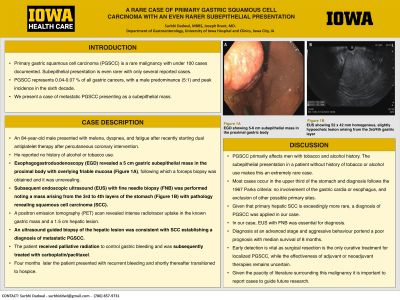
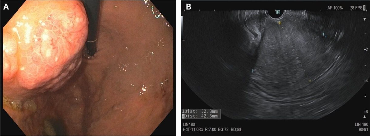
Case Description/Methods: An 84-year-old male presented with melena, dyspnea, and fatigue after recently starting dual antiplatelet therapy after percutaneous coronary intervention. He reported no history of alcohol or tobacco use. Esophagogastroduodenoscopy (EGD) revealed a 5 cm gastric subepithelial mass in the proximal body with overlying friable mucosa (Figure 1A). Forceps biopsy was obtained and unrevealing. Subsequent endoscopic ultrasound (EUS) with fine needle biopsy (FNB) was performed noting a mass arising from the 3rd to 4th layers of the stomach (Figure 1B) with pathology revealing squamous cell carcinoma (SCC). A positron emission tomography (PET) scan revealed intense radiotracer uptake in the known gastric mass and a 1.5 cm hepatic lesion. Ultrasound guided biopsy of the hepatic lesion was consistent with SCC establishing a diagnosis of metastatic PGSCC. The patient received palliative radiation to control gastric bleeding and was subsequently treated with carboplatin/paclitaxel. Four months later the patient presented with recurrent bleeding and shortly thereafter transitioned to hospice.
Discussion: PGSCC primarily affects men with tobacco and alcohol history. The subepithelial presentation in a patient without history of tobacco or alcohol use makes this an extremely rare case. Most cases occur in the upper third of the stomach and diagnosis follows the 1967 Parks criteria: no involvement of the gastric cardia or esophagus, and exclusion of other possible primary sites. Given that primary hepatic SCC is exceedingly more rare a diagnosis of PGSCC was applied in our case. In our case, EUS with FNB was essential for diagnosis. Diagnosis at an advanced stage and aggressive behavior portend a poor prognosis with median survival of 8 months. Early detection is vital as surgical resection is the only curative treatment for localized PGSCC, while the effectiveness of adjuvant or neoadjuvant therapies remains uncertain. Given the paucity of literature surrounding this malignancy it is important to report cases to guide future research.

Disclosures:
Surbhi Dadwal, MBBS1, Joseph Brant, MD2. P1671 - A Rare Case of Primary Gastric Squamous Cell Carcinoma With an Even Rarer Subepithelial Presentation, ACG 2024 Annual Scientific Meeting Abstracts. Philadelphia, PA: American College of Gastroenterology.
1University of Iowa Hospitals & Clinics, Iowa City, IA; 2University of Iowa Hospitals & Clinics, North Liberty, IA
Introduction: Primary gastric squamous cell carcinoma (PGSCC) is a rare malignancy with under 100 cases documented. Subepithelial presentation is even rarer with only several reported cases. PGSCC represents 0.04-0.07 % of all gastric cancers, with a male predominance (5:1) and peak incidence in the sixth decade. We present a case of metastatic PGSCC presenting as a subepithelial mass.
Case Description/Methods: An 84-year-old male presented with melena, dyspnea, and fatigue after recently starting dual antiplatelet therapy after percutaneous coronary intervention. He reported no history of alcohol or tobacco use. Esophagogastroduodenoscopy (EGD) revealed a 5 cm gastric subepithelial mass in the proximal body with overlying friable mucosa (Figure 1A). Forceps biopsy was obtained and unrevealing. Subsequent endoscopic ultrasound (EUS) with fine needle biopsy (FNB) was performed noting a mass arising from the 3rd to 4th layers of the stomach (Figure 1B) with pathology revealing squamous cell carcinoma (SCC). A positron emission tomography (PET) scan revealed intense radiotracer uptake in the known gastric mass and a 1.5 cm hepatic lesion. Ultrasound guided biopsy of the hepatic lesion was consistent with SCC establishing a diagnosis of metastatic PGSCC. The patient received palliative radiation to control gastric bleeding and was subsequently treated with carboplatin/paclitaxel. Four months later the patient presented with recurrent bleeding and shortly thereafter transitioned to hospice.
Discussion: PGSCC primarily affects men with tobacco and alcohol history. The subepithelial presentation in a patient without history of tobacco or alcohol use makes this an extremely rare case. Most cases occur in the upper third of the stomach and diagnosis follows the 1967 Parks criteria: no involvement of the gastric cardia or esophagus, and exclusion of other possible primary sites. Given that primary hepatic SCC is exceedingly more rare a diagnosis of PGSCC was applied in our case. In our case, EUS with FNB was essential for diagnosis. Diagnosis at an advanced stage and aggressive behavior portend a poor prognosis with median survival of 8 months. Early detection is vital as surgical resection is the only curative treatment for localized PGSCC, while the effectiveness of adjuvant or neoadjuvant therapies remains uncertain. Given the paucity of literature surrounding this malignancy it is important to report cases to guide future research.

Figure: Figure 1A. EGD showing 5-6 cm subepithelial mass in the proximal gastric body
Figure 1B. EUS showing 52 x 42 mm homogenous, slightly hypoechoic lesion arising from the 3rd/4th gastric layer
Figure 1B. EUS showing 52 x 42 mm homogenous, slightly hypoechoic lesion arising from the 3rd/4th gastric layer
Disclosures:
Surbhi Dadwal indicated no relevant financial relationships.
Joseph Brant indicated no relevant financial relationships.
Surbhi Dadwal, MBBS1, Joseph Brant, MD2. P1671 - A Rare Case of Primary Gastric Squamous Cell Carcinoma With an Even Rarer Subepithelial Presentation, ACG 2024 Annual Scientific Meeting Abstracts. Philadelphia, PA: American College of Gastroenterology.
