Sunday Poster Session
Category: Stomach
P1674 - PEG, Slug, Bug: Once Seen Never Forgotten
Sunday, October 27, 2024
3:30 PM - 7:00 PM ET
Location: Exhibit Hall E

Has Audio
- AB
Angela Barnes, MD, MBChB
Medical College of Georgia at Augusta University
Augusta, GA
Presenting Author(s)
Award: Presidential Poster Award
Angela Barnes, MD, MBChB1, Phuong Nguyen, MD1, Gurpreet Kaur, MD1, Leonard Little, MD1, Subbaramiah Sridhar, MBBS, MPH2
1Medical College of Georgia at Augusta University, Augusta, GA; 2Augusta University, Augusta, GA
Introduction: Myiasis is an unusual parasitic disease characterized by the invasion of living or necrotic tissues by fly larvae (Diptera family), and can affect both humans and animals. Cutaneous or wound myiasis is relatively uncommon in the United States. We present the third only documented case of PEG stoma myiasis, a rare occurrence involving maggot infestation around the PEG-jejunostomy feeding site.
Case Description/Methods: An 81-year-old male with a history of spinal cord injury and dependent on PEG-Jejunostomy feeding tube and ventilatory support, was admitted to the spinal cord unit. He was unable to communicate in any form. The Gastroenterology Team was consulted for evaluation of his PEG tube site, which was noted to be infested with maggots. The PEG tube had been placed approximately a year previously at another facility. The patient had come from a poorly hygienic living facility. Upon examination, several live maggots were observed at the base of the PEG site, with no evidence of bleeding or sign of gangrene. The PEG tube site was flushed with normal saline, removing approximately 20 live maggots. The tube was eventually changed, effectively clearing the infestation. No more maggots have been noted since then.
Discussion: Cutaneous myiasis is a unique disease that may be missed, particularly in regions where it is uncommon. The etiology of wound myiasis is associated with poor hygiene, where adult female flies lay eggs on or near wounds, leading to rapid larval formation. The larvae then feed directly on the host's tissues, causing the wound to enlarge as they continue to feed. The management of myiasis involves the prompt removal of larvae and appropriate wound care. In our case, the patient's condition was managed successfully with the removal of larvae, simple antisepsis, and a PEG-J tube exchange. The patient's quadriplegia, communication difficulties and prior poor unhygienic living, likely contributed to the infestation. Educating patients and their families about the importance of hygiene and keeping the skin around the PEG tube dry is crucial in preventing this rare occurrence. Healthcare professionals should also remain vigilant in assessing the patients with PEG tubes for any signs of infestation. Our case highlights the importance of the patient and family education regarding preventive measures to avoid exposure to flies and maintain proper hygiene, particularly for individuals with impaired self-awareness and communication.

Disclosures:
Angela Barnes, MD, MBChB1, Phuong Nguyen, MD1, Gurpreet Kaur, MD1, Leonard Little, MD1, Subbaramiah Sridhar, MBBS, MPH2. P1674 - PEG, Slug, Bug: Once Seen Never Forgotten, ACG 2024 Annual Scientific Meeting Abstracts. Philadelphia, PA: American College of Gastroenterology.
Angela Barnes, MD, MBChB1, Phuong Nguyen, MD1, Gurpreet Kaur, MD1, Leonard Little, MD1, Subbaramiah Sridhar, MBBS, MPH2
1Medical College of Georgia at Augusta University, Augusta, GA; 2Augusta University, Augusta, GA
Introduction: Myiasis is an unusual parasitic disease characterized by the invasion of living or necrotic tissues by fly larvae (Diptera family), and can affect both humans and animals. Cutaneous or wound myiasis is relatively uncommon in the United States. We present the third only documented case of PEG stoma myiasis, a rare occurrence involving maggot infestation around the PEG-jejunostomy feeding site.
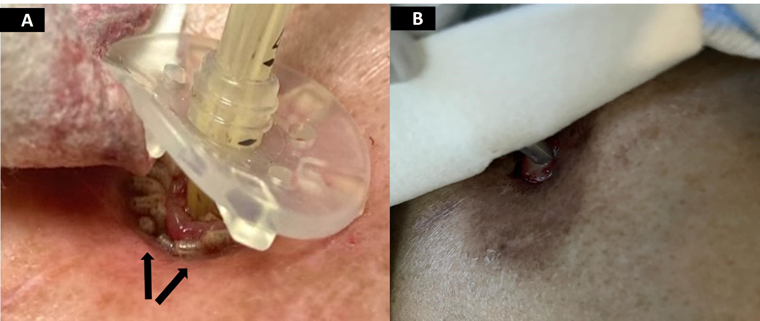
Case Description/Methods: An 81-year-old male with a history of spinal cord injury and dependent on PEG-Jejunostomy feeding tube and ventilatory support, was admitted to the spinal cord unit. He was unable to communicate in any form. The Gastroenterology Team was consulted for evaluation of his PEG tube site, which was noted to be infested with maggots. The PEG tube had been placed approximately a year previously at another facility. The patient had come from a poorly hygienic living facility. Upon examination, several live maggots were observed at the base of the PEG site, with no evidence of bleeding or sign of gangrene. The PEG tube site was flushed with normal saline, removing approximately 20 live maggots. The tube was eventually changed, effectively clearing the infestation. No more maggots have been noted since then.
Discussion: Cutaneous myiasis is a unique disease that may be missed, particularly in regions where it is uncommon. The etiology of wound myiasis is associated with poor hygiene, where adult female flies lay eggs on or near wounds, leading to rapid larval formation. The larvae then feed directly on the host's tissues, causing the wound to enlarge as they continue to feed. The management of myiasis involves the prompt removal of larvae and appropriate wound care. In our case, the patient's condition was managed successfully with the removal of larvae, simple antisepsis, and a PEG-J tube exchange. The patient's quadriplegia, communication difficulties and prior poor unhygienic living, likely contributed to the infestation. Educating patients and their families about the importance of hygiene and keeping the skin around the PEG tube dry is crucial in preventing this rare occurrence. Healthcare professionals should also remain vigilant in assessing the patients with PEG tubes for any signs of infestation. Our case highlights the importance of the patient and family education regarding preventive measures to avoid exposure to flies and maintain proper hygiene, particularly for individuals with impaired self-awareness and communication.

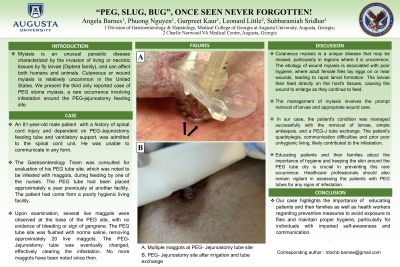
Figure: A) Multiple live maggots at PEG-Jejunostomy tube site
B) PEG-Jejunostomy site after irrigation and tube exchange
B) PEG-Jejunostomy site after irrigation and tube exchange
Disclosures:
Angela Barnes indicated no relevant financial relationships.
Phuong Nguyen indicated no relevant financial relationships.
Gurpreet Kaur indicated no relevant financial relationships.
Leonard Little indicated no relevant financial relationships.
Subbaramiah Sridhar indicated no relevant financial relationships.
Angela Barnes, MD, MBChB1, Phuong Nguyen, MD1, Gurpreet Kaur, MD1, Leonard Little, MD1, Subbaramiah Sridhar, MBBS, MPH2. P1674 - PEG, Slug, Bug: Once Seen Never Forgotten, ACG 2024 Annual Scientific Meeting Abstracts. Philadelphia, PA: American College of Gastroenterology.

