Sunday Poster Session
Category: Interventional Endoscopy
P1097 - Minimally Invasive Marvel: Endoscopic Submucosal Dissection of Colon Polyp - First Case in a Community Hospital
Sunday, October 27, 2024
3:30 PM - 7:00 PM ET
Location: Exhibit Hall E

Has Audio

Pujitha Vallivedu Chennakesavulu, MD, MBBS
Quinnipiac University Frank H. Netter MD School of Medicine / St. Vincent's Medical Center
Bridgeport, CT
Presenting Author(s)
Pujitha Vallivedu Chennakesavulu, MD, MBBS, Sushrut Ingawale, MD, MBBS, Aryak Singh, MD, MBBS, Prabin Sharma, MD
Quinnipiac University Frank H. Netter MD School of Medicine / St. Vincent's Medical Center, Bridgeport, CT
Introduction: Endoscopic resection of gastrointestinal neoplasia and precancerous lesions can be accomplished using endoscopic submucosal dissection (ESD), a highly specialized technique that is performed only in a handful of centers in the United States. ESD is used to remove lesions enbloc and prevent the risk of recurrence. We share our experience of the first case of colon ESD performed at our center.
Case Description/Methods: A 47-year-old female with no significant past medical history underwent an index screening colonoscopy. A 20 mm sessile and multilobulated polyp was noted in the distal rectum and pathology revealed low-grade tubulovillous adenoma. ESD was performed based on characteristics of the lesion, which was found extending a few mm from the dentate line towards the mid-rectum. The polyp was Paris Classification IIa (superficial, elevated). After thermal marking around the polyp, submucosal injection solution was injected with adequate lift of the lesion from muscularis propria. A circumferential incision around the lesion into the submucosa was performed with a Hybrid knife (ERBE). The lesion was then dissected from the underlying deep layers with an electrocautery knife (IT Knife, Hybrid Knife) and retrieved. A 85 X 25 mm lesion was resected. On final pathology, the lesion was tubulovillous adenoma with high-grade dysplasia and the tissue margins were clear of polyp tissue. The patient did well post-procedure and at 6-month follow-up.
Discussion: Endoscopic colon polyp removal can be performed using endoscopic mucosal resection (EMR) or ESD. EMR technique utilizes a snare to remove the polyp after mucosal elevation with a volume expander in a piecemeal fashion. The ESD technique employs a specialized ESD knife to remove the polyp en bloc after dissecting it off the muscularis propria. A metaanalysis has provided the advantage of ESD over EMR in its ability to achieve clear margins with enbloc dissection and eliminate the risk of local polyp recurrence. Setbacks are seen with ESD's longer learning curve, sophisticated skills, prolonged procedure time, and increased risk of bleeding and perforation. ESD is preferred for managing neoplastic lesions due to its superior outcomes, despite technical challenges. Our case highlights the importance of careful case selection and managing colon lesions with ESD. Initial biopsies suggested low-grade dysplasia, but ESD confirmed high-grade with clean margins. ESD prevented the risk of recurrence and the need for multiple procedures in this young patient.

Disclosures:
Pujitha Vallivedu Chennakesavulu, MD, MBBS, Sushrut Ingawale, MD, MBBS, Aryak Singh, MD, MBBS, Prabin Sharma, MD. P1097 - Minimally Invasive Marvel: Endoscopic Submucosal Dissection of Colon Polyp - First Case in a Community Hospital, ACG 2024 Annual Scientific Meeting Abstracts. Philadelphia, PA: American College of Gastroenterology.
Quinnipiac University Frank H. Netter MD School of Medicine / St. Vincent's Medical Center, Bridgeport, CT
Introduction: Endoscopic resection of gastrointestinal neoplasia and precancerous lesions can be accomplished using endoscopic submucosal dissection (ESD), a highly specialized technique that is performed only in a handful of centers in the United States. ESD is used to remove lesions enbloc and prevent the risk of recurrence. We share our experience of the first case of colon ESD performed at our center.
Case Description/Methods: A 47-year-old female with no significant past medical history underwent an index screening colonoscopy. A 20 mm sessile and multilobulated polyp was noted in the distal rectum and pathology revealed low-grade tubulovillous adenoma. ESD was performed based on characteristics of the lesion, which was found extending a few mm from the dentate line towards the mid-rectum. The polyp was Paris Classification IIa (superficial, elevated). After thermal marking around the polyp, submucosal injection solution was injected with adequate lift of the lesion from muscularis propria. A circumferential incision around the lesion into the submucosa was performed with a Hybrid knife (ERBE). The lesion was then dissected from the underlying deep layers with an electrocautery knife (IT Knife, Hybrid Knife) and retrieved. A 85 X 25 mm lesion was resected. On final pathology, the lesion was tubulovillous adenoma with high-grade dysplasia and the tissue margins were clear of polyp tissue. The patient did well post-procedure and at 6-month follow-up.
Discussion: Endoscopic colon polyp removal can be performed using endoscopic mucosal resection (EMR) or ESD. EMR technique utilizes a snare to remove the polyp after mucosal elevation with a volume expander in a piecemeal fashion. The ESD technique employs a specialized ESD knife to remove the polyp en bloc after dissecting it off the muscularis propria. A metaanalysis has provided the advantage of ESD over EMR in its ability to achieve clear margins with enbloc dissection and eliminate the risk of local polyp recurrence. Setbacks are seen with ESD's longer learning curve, sophisticated skills, prolonged procedure time, and increased risk of bleeding and perforation. ESD is preferred for managing neoplastic lesions due to its superior outcomes, despite technical challenges. Our case highlights the importance of careful case selection and managing colon lesions with ESD. Initial biopsies suggested low-grade dysplasia, but ESD confirmed high-grade with clean margins. ESD prevented the risk of recurrence and the need for multiple procedures in this young patient.

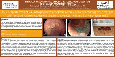
Figure: Image A: Endoscopic view showing sessile and multi-lobular polyp in the mid rectum, Image B: Endoscopic view of the rectum after ESD,
Image C: Gross view of resected polyp - 85 X 25 mm
Image C: Gross view of resected polyp - 85 X 25 mm
Disclosures:
Pujitha Vallivedu Chennakesavulu indicated no relevant financial relationships.
Sushrut Ingawale indicated no relevant financial relationships.
Aryak Singh indicated no relevant financial relationships.
Prabin Sharma indicated no relevant financial relationships.
Pujitha Vallivedu Chennakesavulu, MD, MBBS, Sushrut Ingawale, MD, MBBS, Aryak Singh, MD, MBBS, Prabin Sharma, MD. P1097 - Minimally Invasive Marvel: Endoscopic Submucosal Dissection of Colon Polyp - First Case in a Community Hospital, ACG 2024 Annual Scientific Meeting Abstracts. Philadelphia, PA: American College of Gastroenterology.
