Sunday Poster Session
Category: GI Bleeding
P0801 - Unveiling Dieulafoy’s Lesion: Concealed Beneath a Clot in Sigmoid Colon
Sunday, October 27, 2024
3:30 PM - 7:00 PM ET
Location: Exhibit Hall E

Has Audio
.jpg)
Narinderjeet Kaur, MD, MS
SUNY Downstate Medical Center
Brooklyn, NY
Presenting Author(s)
Narinderjeet Kaur, MD, MS, Jonathan Lo, MD, Anwar Uddin, MD, Benjamin Silver, MD, Yaniuska Lescaille, MD, MPH, Qi Yu, MD, MPH
SUNY Downstate Medical Center, Brooklyn, NY
Introduction: Dieulafoy’s lesion (DL) is an abnormally dilated submucosal vessel that can present as an abrupt gastrointestinal bleed (GIB). These lesions are commonly found in the lesser curvature of the stomach or esophagus and are rare in the colon. Gastrointestinal bleeds associated with these lesions can range from mild, recurrent to life-threatening hemorrhages. Their small size makes it difficult to diagnose. DL is a rarer cause of lower GIB compared to common etiologies like diverticulosis, hemorrhoids, and angiodysplasia. We present a case of a 66-year-old female presenting with bright red blood per rectum caused by sigmoid colon DL.
Case Description/Methods: A 66-year-old female presented to the hospital with lower abdominal pain and bright red blood per rectum (BRBPR). Her medical history was significant for hypertension, diabetes, hypothyroidism, atrial fibrillation, diverticulosis, and ventral hernia that was post-repair. The patient had a similar episode of BRBPR ten years ago, and a colonoscopy performed at that time was unrevealing. Upon presentation, vital signs were significant for blood pressure of 152/86 mmHg and heart rate of 97. The digital rectal exam was remarkable for maroon-colored stool. Labs revealed a hemoglobin 9.0mg/dL, which was similar to her baseline. A colonoscopy was performed, and a small adherent blood clot was observed in the sigmoid colon (Figure 1 A). The clot was washed, and it revealed a punctate area of active blood oozing surrounded by normal mucosa. To stop the active bleeding, two hemostatic clips were placed with successful hemostasis. The colonoscopy was unremarkable otherwise.
Discussion: Endoscopy carries a 70% success rate of identifying a DL lesion via direct endoscopic visualization. The lack of mucosal defect at the edge of the lesion results in a diagnostic dilemma when a vessel is too small to be visualized on an endoscopy, such as a vessel size of 1 to 5 mm with a pulsatile or an actively bleeding vessel. Therapeutic endoscopic hemostasis is the mainstay of DL management as performed in our case. Angiography by interventional radiology (IR) can be a valuable alternative for inaccessible lesions, and surgical intervention is the last resort for the management of lesions that fail to respond to endoscopic and IR interventions.

Disclosures:
Narinderjeet Kaur, MD, MS, Jonathan Lo, MD, Anwar Uddin, MD, Benjamin Silver, MD, Yaniuska Lescaille, MD, MPH, Qi Yu, MD, MPH. P0801 - Unveiling Dieulafoy’s Lesion: Concealed Beneath a Clot in Sigmoid Colon, ACG 2024 Annual Scientific Meeting Abstracts. Philadelphia, PA: American College of Gastroenterology.
SUNY Downstate Medical Center, Brooklyn, NY
Introduction: Dieulafoy’s lesion (DL) is an abnormally dilated submucosal vessel that can present as an abrupt gastrointestinal bleed (GIB). These lesions are commonly found in the lesser curvature of the stomach or esophagus and are rare in the colon. Gastrointestinal bleeds associated with these lesions can range from mild, recurrent to life-threatening hemorrhages. Their small size makes it difficult to diagnose. DL is a rarer cause of lower GIB compared to common etiologies like diverticulosis, hemorrhoids, and angiodysplasia. We present a case of a 66-year-old female presenting with bright red blood per rectum caused by sigmoid colon DL.
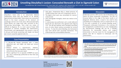
Case Description/Methods: A 66-year-old female presented to the hospital with lower abdominal pain and bright red blood per rectum (BRBPR). Her medical history was significant for hypertension, diabetes, hypothyroidism, atrial fibrillation, diverticulosis, and ventral hernia that was post-repair. The patient had a similar episode of BRBPR ten years ago, and a colonoscopy performed at that time was unrevealing. Upon presentation, vital signs were significant for blood pressure of 152/86 mmHg and heart rate of 97. The digital rectal exam was remarkable for maroon-colored stool. Labs revealed a hemoglobin 9.0mg/dL, which was similar to her baseline. A colonoscopy was performed, and a small adherent blood clot was observed in the sigmoid colon (Figure 1 A). The clot was washed, and it revealed a punctate area of active blood oozing surrounded by normal mucosa. To stop the active bleeding, two hemostatic clips were placed with successful hemostasis. The colonoscopy was unremarkable otherwise.
Discussion: Endoscopy carries a 70% success rate of identifying a DL lesion via direct endoscopic visualization. The lack of mucosal defect at the edge of the lesion results in a diagnostic dilemma when a vessel is too small to be visualized on an endoscopy, such as a vessel size of 1 to 5 mm with a pulsatile or an actively bleeding vessel. Therapeutic endoscopic hemostasis is the mainstay of DL management as performed in our case. Angiography by interventional radiology (IR) can be a valuable alternative for inaccessible lesions, and surgical intervention is the last resort for the management of lesions that fail to respond to endoscopic and IR interventions.

Figure: Figure 1A) Adherent clot in the sigmoid colon (yellow arrows). Figure 1B) Punctate area of active oozing. Figure 1C) Hemostatic clip successfully deployed with complete hemostasis.
Disclosures:
Narinderjeet Kaur indicated no relevant financial relationships.
Jonathan Lo indicated no relevant financial relationships.
Anwar Uddin indicated no relevant financial relationships.
Benjamin Silver indicated no relevant financial relationships.
Yaniuska Lescaille indicated no relevant financial relationships.
Qi Yu indicated no relevant financial relationships.
Narinderjeet Kaur, MD, MS, Jonathan Lo, MD, Anwar Uddin, MD, Benjamin Silver, MD, Yaniuska Lescaille, MD, MPH, Qi Yu, MD, MPH. P0801 - Unveiling Dieulafoy’s Lesion: Concealed Beneath a Clot in Sigmoid Colon, ACG 2024 Annual Scientific Meeting Abstracts. Philadelphia, PA: American College of Gastroenterology.
