Sunday Poster Session
Category: Esophagus
P0594 - Dysphagia As a Presenting Symptom of An Aggressive Adenocarcinoma of Lung In a Female
Sunday, October 27, 2024
3:30 PM - 7:00 PM ET
Location: Exhibit Hall E

Has Audio

Neil Gambhir, DO
Lenox Hill Hospital, Northwell Health
New York, NY
Presenting Author(s)
Neil Gambhir, DO1, Emily A. Rodriguez, DO1, Patricia Lin, MS1, Moein Bayat Mokhtari, MD1, Jonathan Jeong, DO1, Samantha Rubin, DO1, Thida Aye, MD, MPH.2, Wan Lam, MD2
1Lenox Hill Hospital, Northwell Health, New York, NY; 2Lenox Hill, Northwell Health, New York, NY
Introduction: Lung cancer is the leading cause of cancer deaths in the United States and the second most prevalent cancer overall. Though typically associated with respiratory symptoms, dysphagia can be a rare initial presentation in 1-2% of cases. Herein, we detail the case of a 53-year-old Asian female with no smoking history who presented with abrupt dysphagia found to have a large and aggressive adenocarcinoma of the lung.
Case Description/Methods: The patient is a 53-year-old Asian female without significant past medical history who presented with progressive sudden dysphagia with a week's duration. Before admission, the patient underwent an esophagogastroduodenoscopy revealing external compression of the esophagus with X-ray demonstrating a 3-cm mass in the right lower lung lobe
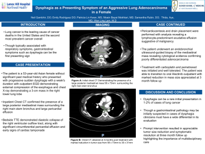
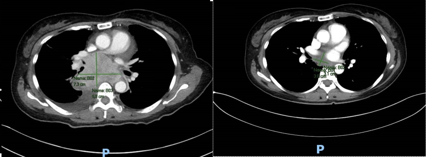
Upon admission, the patient was hemodynamically stable with lab work significant for mild hyponatremia and anion gap acidosis indicative of starvation ketosis. Initial chest CT confirmed the presence of a large posterior mediastinal mass (7.3 cm), surrounding the right main stem bronchus and large pericardial effusion.
Bedside transthoracic echocardiogram demonstrated diastolic collapse of the right ventricular outflow tract, along with significant circumferential pericardial effusion and early signs of cardiac tamponade. The patient underwent pericardiocentesis and drain placement, which evacuated 1000 cc of straw-colored fluid. Analysis of the pericardial fluid revealed a lymphocyte-predominant exudative pericardial effusion suggestive of malignancy. The patient then underwent an endobronchial ultrasound-guided biopsy of the mediastinal mass, which revealed cytological evidence confirming poorly differentiated adenocarcinoma
Inpatient treatment with carboplatin and pemetrexed effectively debulked the tumor and was well tolerated by the patient. They were successfully transitioned to an oral regimen with Alectinib. Latest imaging conducted at 3 months post-discharge showed a remarkable improvement, with near complete resolution of the right-sided lung mass and a notable reduction in the size of the mediastinal mass.
Discussion: While lung cancer presents mainly with respiratory symptoms, atypical presentations including that of dysphagia in an otherwise healthy patient should not be disregarded. Early recognition of malignancy is crucial for prompt intervention and follow-up to achieve the best outcomes in these patients.

Disclosures:
Neil Gambhir, DO1, Emily A. Rodriguez, DO1, Patricia Lin, MS1, Moein Bayat Mokhtari, MD1, Jonathan Jeong, DO1, Samantha Rubin, DO1, Thida Aye, MD, MPH.2, Wan Lam, MD2. P0594 - Dysphagia As a Presenting Symptom of An Aggressive Adenocarcinoma of Lung In a Female, ACG 2024 Annual Scientific Meeting Abstracts. Philadelphia, PA: American College of Gastroenterology.
1Lenox Hill Hospital, Northwell Health, New York, NY; 2Lenox Hill, Northwell Health, New York, NY
Introduction: Lung cancer is the leading cause of cancer deaths in the United States and the second most prevalent cancer overall. Though typically associated with respiratory symptoms, dysphagia can be a rare initial presentation in 1-2% of cases. Herein, we detail the case of a 53-year-old Asian female with no smoking history who presented with abrupt dysphagia found to have a large and aggressive adenocarcinoma of the lung.
Case Description/Methods: The patient is a 53-year-old Asian female without significant past medical history who presented with progressive sudden dysphagia with a week's duration. Before admission, the patient underwent an esophagogastroduodenoscopy revealing external compression of the esophagus with X-ray demonstrating a 3-cm mass in the right lower lung lobe
Upon admission, the patient was hemodynamically stable with lab work significant for mild hyponatremia and anion gap acidosis indicative of starvation ketosis. Initial chest CT confirmed the presence of a large posterior mediastinal mass (7.3 cm), surrounding the right main stem bronchus and large pericardial effusion.
Bedside transthoracic echocardiogram demonstrated diastolic collapse of the right ventricular outflow tract, along with significant circumferential pericardial effusion and early signs of cardiac tamponade. The patient underwent pericardiocentesis and drain placement, which evacuated 1000 cc of straw-colored fluid. Analysis of the pericardial fluid revealed a lymphocyte-predominant exudative pericardial effusion suggestive of malignancy. The patient then underwent an endobronchial ultrasound-guided biopsy of the mediastinal mass, which revealed cytological evidence confirming poorly differentiated adenocarcinoma
Inpatient treatment with carboplatin and pemetrexed effectively debulked the tumor and was well tolerated by the patient. They were successfully transitioned to an oral regimen with Alectinib. Latest imaging conducted at 3 months post-discharge showed a remarkable improvement, with near complete resolution of the right-sided lung mass and a notable reduction in the size of the mediastinal mass.
Discussion: While lung cancer presents mainly with respiratory symptoms, atypical presentations including that of dysphagia in an otherwise healthy patient should not be disregarded. Early recognition of malignancy is crucial for prompt intervention and follow-up to achieve the best outcomes in these patients.

Figure: Near complete resolution of the right-sided lung mass is appreciated with a notable reduction in the size of the mediastinal mass. Specifically, the measurements decreased from the initial size of 58 x 73mm to 30 x 31mm as demonstrated by the image on the left (initial CT scan) and the image on the right (CT at 3 month follow-up)
Disclosures:
Neil Gambhir indicated no relevant financial relationships.
Emily Rodriguez indicated no relevant financial relationships.
Patricia Lin indicated no relevant financial relationships.
Moein Bayat Mokhtari indicated no relevant financial relationships.
Jonathan Jeong indicated no relevant financial relationships.
Samantha Rubin indicated no relevant financial relationships.
Thida Aye indicated no relevant financial relationships.
Wan Lam indicated no relevant financial relationships.
Neil Gambhir, DO1, Emily A. Rodriguez, DO1, Patricia Lin, MS1, Moein Bayat Mokhtari, MD1, Jonathan Jeong, DO1, Samantha Rubin, DO1, Thida Aye, MD, MPH.2, Wan Lam, MD2. P0594 - Dysphagia As a Presenting Symptom of An Aggressive Adenocarcinoma of Lung In a Female, ACG 2024 Annual Scientific Meeting Abstracts. Philadelphia, PA: American College of Gastroenterology.
