Sunday Poster Session
Category: Esophagus
P0528 - Spinal Fracture: An Uncommon Primary Presentation of Esophageal Adenocarcinoma
Sunday, October 27, 2024
3:30 PM - 7:00 PM ET
Location: Exhibit Hall E

Has Audio
- SM
Swayamdipto Misra, MD
UT Health Science Center
Tyler, TX
Presenting Author(s)
Swayamdipto Misra, MD1, Layth Alzubaidy, MD2, Bolarinwa Olusola, MD3
1UT Health Science Center, Tyler, TX; 2University of Texas Health Science Center, Tyler, TX; 3University of Texas Health East Texas Physicians, Tyler, TX
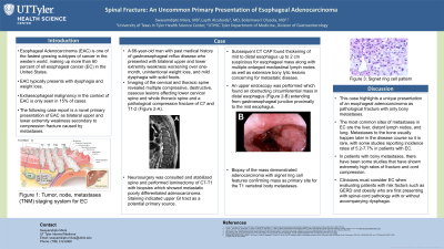
Introduction: Esophageal Adenocarcinoma (EAC) is one of the fastest-growing subtypes of cancer in the Western world, making up more than 60 percent of all esophageal cancer (EC) in the United States. It typically presents with dysphagia and weight loss. Extraesophageal presentation of EAC is seen in nearly 15% of EAC cases. Here we present a case of a pathological fracture as the initial clinical manifestation of an advanced EAC.
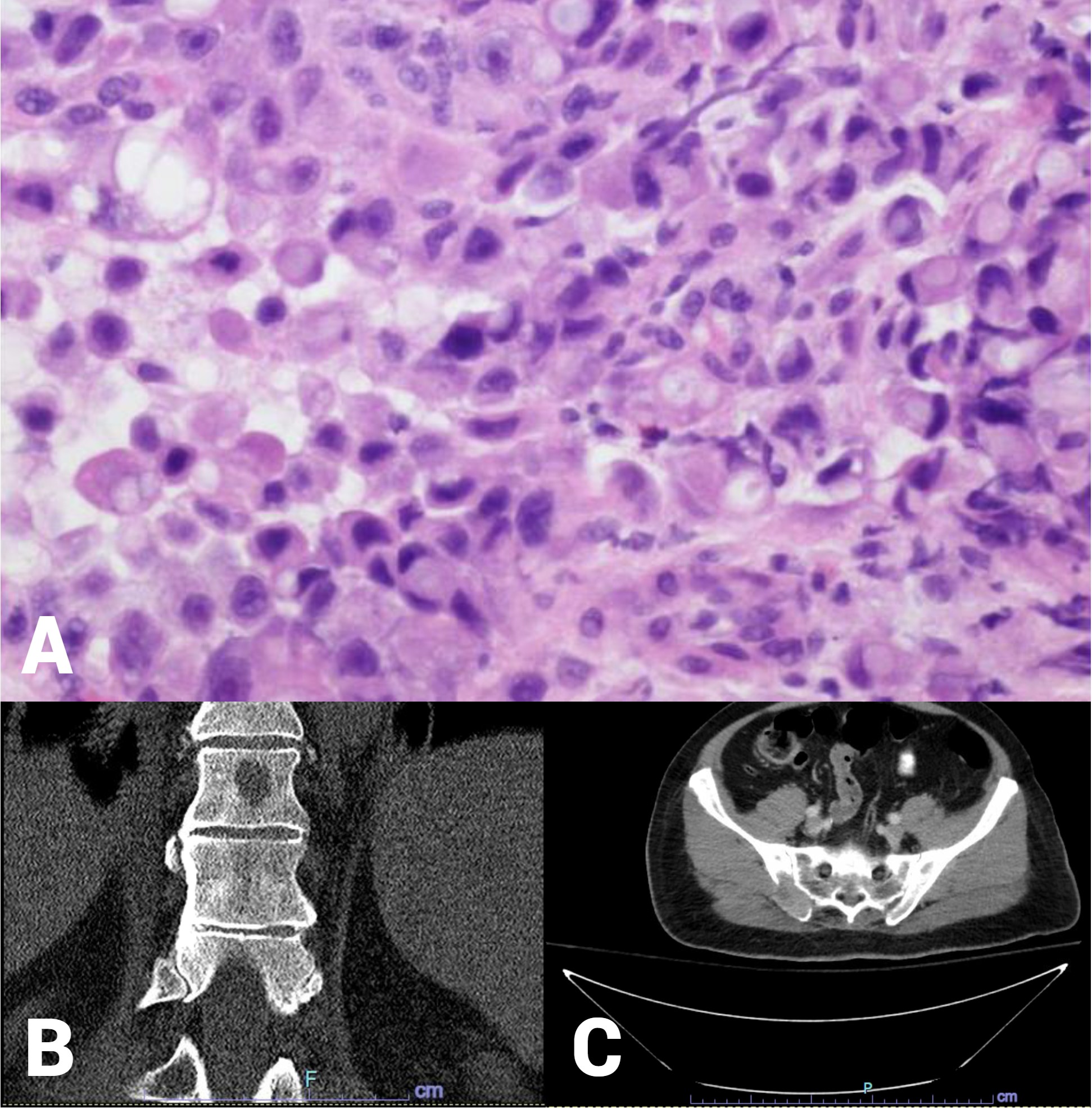
Case Description/Methods: A 66-year-old man with a past medical history of gastroesophageal reflux disease presented with bilateral upper and lower extremity weakness worsening over one month, unintentional weight loss, and mild dysphagia with solid foods. Computed Topography (CT) of the cervical spine revealed diffuse osseous metastatic disease throughout the thoracic spine and pathologic compression fracture at T1. Neurosurgery service was consulted and stabilized spine and laminectomy of C7-T1 with biopsies which showed metastatic poorly differentiated adenocarcinoma. Staining indicated the upper GI tract as a potential primary source. Subsequent CT of the abdomen and pelvis revealed numerous scattered lytic lesions involving the thoracolumbar spine, ribs, sternum, left scapula, left 1st rib head, and left C7 facet (Figure 1B-C). In addition, it showed thickening of mid to distal esophagus up to 2 cm suspicious for esophageal mass along with multiple enlarged mediastinal lymph nodes. Upper endoscopy found an obstructing circumferential mass in the distal esophagus extending from the gastroesophageal junction proximally to the mid-esophagus. Biopsy of the mass showed adenocarcinoma with signet ring cell features confirming mass as the primary site for the T1 vertebral body metastasis (figure 1A).
Discussion: This case highlights a unique presentation of an esophageal adenocarcinoma as a pathological fracture of the spine. While the patient in this case had mild dysphagia, his primary presenting symptom was neurological as evidenced by upper and lower extremity weakness. Adenocarcinomas most frequently metastasize to intraabdominal sites such as the liver and peritoneum. However, this case demonstrated no merely mediastinal lymphadenopathy and diffuse osseous metastasis, which is an uncommon behavior of EAC.

Disclosures:
Swayamdipto Misra, MD1, Layth Alzubaidy, MD2, Bolarinwa Olusola, MD3. P0528 - Spinal Fracture: An Uncommon Primary Presentation of Esophageal Adenocarcinoma, ACG 2024 Annual Scientific Meeting Abstracts. Philadelphia, PA: American College of Gastroenterology.
1UT Health Science Center, Tyler, TX; 2University of Texas Health Science Center, Tyler, TX; 3University of Texas Health East Texas Physicians, Tyler, TX
Introduction: Esophageal Adenocarcinoma (EAC) is one of the fastest-growing subtypes of cancer in the Western world, making up more than 60 percent of all esophageal cancer (EC) in the United States. It typically presents with dysphagia and weight loss. Extraesophageal presentation of EAC is seen in nearly 15% of EAC cases. Here we present a case of a pathological fracture as the initial clinical manifestation of an advanced EAC.
Case Description/Methods: A 66-year-old man with a past medical history of gastroesophageal reflux disease presented with bilateral upper and lower extremity weakness worsening over one month, unintentional weight loss, and mild dysphagia with solid foods. Computed Topography (CT) of the cervical spine revealed diffuse osseous metastatic disease throughout the thoracic spine and pathologic compression fracture at T1. Neurosurgery service was consulted and stabilized spine and laminectomy of C7-T1 with biopsies which showed metastatic poorly differentiated adenocarcinoma. Staining indicated the upper GI tract as a potential primary source. Subsequent CT of the abdomen and pelvis revealed numerous scattered lytic lesions involving the thoracolumbar spine, ribs, sternum, left scapula, left 1st rib head, and left C7 facet (Figure 1B-C). In addition, it showed thickening of mid to distal esophagus up to 2 cm suspicious for esophageal mass along with multiple enlarged mediastinal lymph nodes. Upper endoscopy found an obstructing circumferential mass in the distal esophagus extending from the gastroesophageal junction proximally to the mid-esophagus. Biopsy of the mass showed adenocarcinoma with signet ring cell features confirming mass as the primary site for the T1 vertebral body metastasis (figure 1A).
Discussion: This case highlights a unique presentation of an esophageal adenocarcinoma as a pathological fracture of the spine. While the patient in this case had mild dysphagia, his primary presenting symptom was neurological as evidenced by upper and lower extremity weakness. Adenocarcinomas most frequently metastasize to intraabdominal sites such as the liver and peritoneum. However, this case demonstrated no merely mediastinal lymphadenopathy and diffuse osseous metastasis, which is an uncommon behavior of EAC.

Figure: Figure 1: (A) histologic image (H&E 400x) showing poorly differentiated adenocarcinoma. (B) example of osseous metastasis to the thoracolumbar region and iliac crest (C).
Disclosures:
Swayamdipto Misra indicated no relevant financial relationships.
Layth Alzubaidy indicated no relevant financial relationships.
Bolarinwa Olusola indicated no relevant financial relationships.
Swayamdipto Misra, MD1, Layth Alzubaidy, MD2, Bolarinwa Olusola, MD3. P0528 - Spinal Fracture: An Uncommon Primary Presentation of Esophageal Adenocarcinoma, ACG 2024 Annual Scientific Meeting Abstracts. Philadelphia, PA: American College of Gastroenterology.
